Carlos E Marroquin, Pam Gibson, Jon Boyson and Mark Fung
Sean M Wrenn, Pamela C Gibson, Donna-Sue Hain, Sarah Harm, Paulette B Hammond, Diti H Shah, Susan E Hillyard, Mark Fung and Carlos E Marroquin
DOI: 10.4172/2476-1966.1000101
Introduction: Hemolytic uremic syndrome (HUS) following kidney transplantation is a devastating complication that may result in substantial morbidity and allograft loss. While calcineurin inhibitor-induced HUS has been well described, anti-donor specific antibody production may be an alternative pathway in the pathogenesis of HUS. We present a case of HUS following kidney transplantation, and will present evidence that donor specific antibodies (DSA) may be a factor in platelet activation and end-organ injury resulting in post-transplant HUS. Case Description: A 66-year-old female with diabetic nephropathy underwent a deceased donor renal transplant with a 5 HLA mismatched kidney. Her immediate course was uneventful with normalization of her creatinine (Cr). She was re-admitted with rising Cr, oliguria, proteinuria, anaemia and thrombocytopenia. A peripheral smear revealed schistocytes, haptoglobin levels were depleted and an allograft biopsy was performed that was suggestive of thrombotic microangiopathy (TMA) with equivocal findings for AMR. Her ADAMTS 13 activity was 103% (normal). She concurrently developed a substantial de novo DSA burden. She underwent therapeutic plasma-pheresis, conversion from tacrolimus to cyclosporine, and received rituximab therapy. She had a complete clinical resolution and remains off of dialysis. Her laboratory markers improved and her antibody titers decreased. Discussion: Post-transplant HUS requires immediate recognition and treatment. This clinical course suggests DSA may be involved in an alternative mechanism of platelet activation leading to HUS and renal insult. Review of the literature suggests this is a rare cause of HUS and we postulate may be under-diagnosed in the transplant population and requires further study.
The coexistence of Graves’ disease (GD) and inflammatory bowel disease (IBD), particularly Crohn’s disease (CD) and ulcerative colitis (UC), has not been well documented. Therefore, this report reviews the literature regarding coexisting IBD and GD. Reported cases of concomitant IBD and GD are rare; 16 cases of concomitant UC and GD and 3 cases of concomitant CD and GD were found. Among the 19 reported cases of concomitant IBD and GD, IBD developed before GD in 8 (42.1%) cases, GD developed before IBD in 9 (47.4%) cases, and both conditions coexisted in 2 (10.5%) cases. Therefore, there was no evidence for a tendency of a preceding disease between IBD and GD. The interval between diagnoses of IBD and GD varied from 0 year to 20 years. Furthermore, there was no evidence indicating that patients with concomitant IBD and GD had poorer prognoses than those with IBD but without GD.
Malgorzata Kloc, Xian C Li and Rafik M Ghobrial
Holthaus KB, Spisni E and Alibardi L
Previous in-vivo studies have isolated and identified peptides with typical molecular anti-microbial characteristics in reptiles. In the present study we have tested the putative antimicrobial action of a lizard cathelicidin and of a turtle beta-defensin using the broth microdilution assay on Gram positive and Gram negative bacteria. The addition of the peptides at concentrations indicatively ranging between 0.05-1.9 mg/ml (cathelicidin) and 0.69-4.14 mg/ml (betadefensin) inhibited bacterial growth after 3 hours of incubation as determined by their MIC and IC50 values. Due to the poor solubility and the medium interference the real concentration of the delivered peptides to the bacterial cultures was uncertain. The qualitative evaluation of the anti-microbial damage after treatment with the peptides was done under the electron microscope that showed some alteration and rupture in the plasma membrane, lowering of the ribosomes, swelling and clumping in nucleoid region of Gram negative (E. coli) and Gram positive (S. aureus) bacteria. Immunogold labeling against the two peptides indicated that the peptides were localized not only on the plasma membrane and in cytoplasm of the treated bacteria, but also in the nucleoid region and its protein scaffold. The present ultrastructural study suggests that these peptides operate a cellular damage initially on the plasma membrane but further also in the ribosomes and on the DNA or its associated proteins.
Maria Biba, Theodora Keramitsoglou, Dimitris Goukos, Marigoula Varla-Leftherioti, Konstantinos Pantos, George Koumantakis, Antonis Makrigiannakis and Ioannis E. Messinis
Objective: Interleukin-1 beta (IL-1β) and Interleukin-6 (IL-6) are pro-inflammatory cytokines involved in the mother-embryo interaction during blastocyst adhesion and invasion into the endometrium. IL-1β is also considered as a first signal delivered from blastocyst to the endometrium to influence uterus receptivity. The aim of the present prospective, non-randomized study was to explore whether the measurement of IL-1β and IL-6 secretion by blastocysts could serve as a non-invasive method to predict blastocyst implantation competence following in vitro fertilization (IVF). Methods: IL-1β and IL-6 were measured in the supernatant culture media of 683 blastocysts transferred into 245 women following IVF cycles, and their levels were correlated with implantation and pregnancy rates per transfer. Measurements were performed using a Luminex 200 and commercially available interleukin kits. Statistical analyses were performed using GraphPad Prism 5 software, and a probability of p<0.05 was used to indicate a significant difference. Results: IL-1β was detected in 26.5% of the blastocyst supernatants (179/683), with a mean value 0.099 pg/ml, and IL-6 was detected in 20.35% of blastocysts (139/683), with a mean value of 0.046 pg/ml. Cytokine levels showed no correlation with blastocyst quality or developmental stage. The mean values of IL-1β and IL-6 in the implanted blastocysts were 0.073 and 0.036 pg/ml, respectively. In the non-implanted blastocysts, the corresponding values were 0.0141 and 0.060 pg/ml. The pregnancy and implantation rates in women with detectable IL-1β levels (pregnancy rate (PR): 56.3%; implantation rate (IR): 21.8%) and non-detectable IL-1β levels (PR: 60.23%; IR: 28.37%) and in women with detectable IL-6 levels (PR: 58%; IR: 25%) and non-detectable IL-6 levels (PR: 59.5%; IR: 26.83%) were not significantly different. Conclusion: Blastocysts secrete IL-1β, and to a lesser extent, IL-6. No significant differences in implantation or pregnancy outcomes were identified between patients with detectable and undetectable IL-1β and IL-6 levels. Therefore, the non-invasive measurement of IL-1β and IL-6 secreted by blastocysts prior to transfer should not be considered a useful biomarker of blastocyst development and implantation competence.
Zhichao Fan and Wei Liu
DOI: 10.4172/2476-1966.1000106
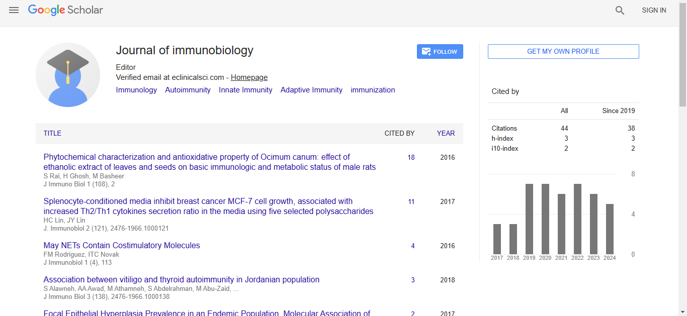
Journal of Immunobiology received 34 citations as per Google Scholar report

 Spanish
  Chinese
  Russian
  German
  French
  Japanese
  Portuguese
  Hindi
Hindi 