Brief Report - (2025) Volume 10, Issue 1
Received: 01-Mar-2025, Manuscript No. jib-25-168749;
Editor assigned: 03-Mar-2025, Pre QC No. P-168749;
Reviewed: 15-Mar-2025, QC No. Q-168749;
Revised: 20-Mar-2025, Manuscript No. R-168749;
Published:
27-Mar-2025
, DOI: 10.37421/2476-1966.2025.10.262
Citation: Mirzaei, Weller. “Antibody-mediated Modulation of the Tumor Microenvironment: Mechanistic Insights and Clinical Outcomes.” J Immuno Biol 10 (2025): 262.
Copyright: © 2025 Mirzaei W. This is an open-access article distributed under the terms of the Creative Commons Attribution License which permits unrestricted use, distribution and reproduction in any medium, provided the original author and source are credited.
The TME is composed of a heterogeneous mix of immune and non-immune cells that interact through direct contact and soluble factors. These include cytotoxic T cells, regulatory T cells (Tregs), myeloid-derived suppressor cells, tumor-associated macrophages, cancer-associated fibroblasts, endothelial cells, and more. In the immunosuppressive TME, cytotoxic immune cells are often rendered dysfunctional, while immunosuppressive cells and signals dominate, contributing to immune evasion and tumor growth. Modulating these dynamics using monoclonal antibodies represents a pivotal strategy in reactivating the immune system and restoring immunosurveillance [2].
One of the most successful applications of monoclonal antibodies in oncology is their use in immune checkpoint blockade. As described in the previous discussion, antibodies such as anti-PD-1 (nivolumab, pembrolizumab) and anti-CTLA-4 (ipilimumab) function by inhibiting negative regulatory signals that limit T cell activation. In the context of the TME, these antibodies help rescue Tumor-Infiltrating Lymphocytes (TILs) from functional exhaustion, increasing their proliferation, cytokine production, and cytotoxic activity. Clinical studies have shown that tumors with higher baseline levels of PD-L1 expression or greater TIL density are more likely to respond to checkpoint inhibitors, underscoring the critical role of the TME in determining therapeutic efficacy. In addition to checkpoint inhibitors, other antibody-based strategies directly target immunosuppressive cells in the TME. Antibodies targeting colony-stimulating factor 1 receptor (CSF1R), such as emactuzumab and cabiralizumab, deplete or reprogram TAMs, shifting the balance toward an anti-tumor M1-like phenotype. Similarly, MDSCs are targeted using anti-CD33 antibodies (e.g., gemtuzumab ozogamicin), which help reduce their immunosuppressive burden and improve T cell responses [3].
Tumor heterogeneity and immune evasion remain challenges in optimizing antibody-based modulation of the TME. Many tumors develop resistance to single-agent immunotherapy by upregulating alternative immune checkpoints, altering antigen presentation machinery, or recruiting suppressive immune cells. To counter these adaptations, combination strategies are being pursued. These include combinations of checkpoint inhibitors (dual blockade), checkpoint inhibitors with targeted therapies, and antibodies with radiation or chemotherapy. Combinations targeting both the immune and stromal compartments of the TME have shown particular promise in enhancing immune infiltration and overcoming resistance [4,5].
Google Scholar Cross Ref Indexed at
Google Scholar Cross Ref Indexed at
Google Scholar Cross Ref Indexed at
Google Scholar Cross Ref Indexed at
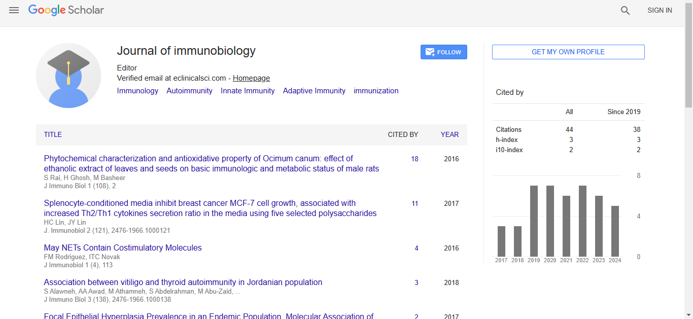
Journal of Immunobiology received 34 citations as per Google Scholar report