Mini Review - (2022) Volume 12, Issue 6
Received: 15-Nov-2022, Manuscript No. jttr-23-85347;
Editor assigned: 17-Nov-2022, Pre QC No. P-85347;
Reviewed: 22-Nov-2022, QC No. Q-85347;
Revised: 02-Dec-2022, Manuscript No. R-85347;
Published:
09-Dec-2022
, DOI: 10.37421/2161-0991.2022.12.222
Citation: Sallinen, Ville. “A Functioning and Vascularized Human Brain Organoids.” J Transplant Technol Res 12 (2022): 222.
Copyright: © 2022 Sallinen V. This is an open-access article distributed under the terms of the Creative Commons Attribution License, which permits unrestricted use, distribution, and reproduction in any medium, provided the original author and source are credited.
An unprecedented opportunity exists to simulate human brain development and disease by dividing human pluripotent stem cells into tiny brainlike organoids. We developed a technique for transplanting human brain organoids into adult mouse brains in order to provide a vascularized and functional in vivo model of brain organoids. Organoid joins showed moderate neuronal separation and development, gliogenesis, combination of microglia and development of axons to various areas of the host cerebrum. Two-photon in vivo imaging revealed the grafts' functional neuronal networks and blood vessels. Finally, optogenetics and in vivo extracellular recording revealed intragraft neuronal activity and suggested functional synaptic connectivity between the host and the graft. It's possible that disease modeling under physiological conditions will be made easier by combining human neural organoids with an in vivo physiological environment in the animal brain.
Brain death • Humans • Kidney • Pig • Xenotransplantation
Is there new information gleaned from this experiment and does it pave the way for future clinical trials? Although there was very little information provided, the following points are important to note: i) The pig kidney is predicted to function immediately by numerous in vivo studies on nonhuman primates. ii) Numerous in vitro studies have predicted that a GTKO pig kidney transplanted into a human subject would not be rejected within the first few days. iii) A triple-knockout (TKO) pig kidney transplant would have been more appropriate because GTKO kidneys are not ideal for clinical transplantation. iv) Transplanting a "thymokidney" without a pre-transplant conditioning treatment and several months of follow-up was pointless. v) It is difficult to determine whether the graft's function was sufficient to support life because the native kidneys were retained. vi) Although it is hoped that it will be published in a peerreviewed medical journal, the fact that the experiment was only announced to the media suggests that it was primarily carried out to draw attention to the enormous potential of xenotransplantation (and/or possibly to NYU). The experiment was successful in this regard.
Over the past few decades, significant advancements in immunosuppressive treatment, surgical techniques and post-transplant complications have significantly improved renal transplant outcomes. When transplant outcomes from living and deceased donors are compared, these accomplishments have not, however, resulted in improvements that are comparable. Acceptance of suboptimal donors has increased as a result of the high demand for donor kidneys. The number of renal transplants has greatly increased as a result of the use of brain-dead patients as organ donors. Sadly, brain death, a physiologically abnormal state, has a negative impact on transplant outcomes. Potential grafts are already damaged prior to retrieval and preservation, as evidenced by the decreased viability of transplanted kidneys derived from brain-dead donors. We provide an overview of the current understanding of the (patho) physiological effects of brain death and how they relate to the outcome of renal transplants in this review. In addition, a number of options for therapeutic intervention during the donor's brain death are discussed with the aim of increasing organ viability and transplant success.
The orthotopic liver transplants from deceased donors that were carried out in Finland between June 2004 and December 2017 were included in the Finnish Cohort and they were followed up until either death or retransplantation in October 2020. The donor's medical records and the Finnish Transplant Registry were used to extract the data. International organ exchanges were not included in the study. The same group of transplant surgeons from Helsinki Transplantation and Liver Surgery Unit procured all of the included organs within Finland. All transplants in Finland were carried out at Helsinki University Hospital. During the study period, all liver grafts were donated following brain death (DBD). US Cohort The Scientific Registry of Transplant Recipients (SRTR) provided the data for this study. The Organ Procurement and Transplantation Network (OPTN) members who submitted data on all donors, wait-list candidates and transplant recipients in the United States are included in the SRTR data system. The activities of the OPTN and SRTR contractors are overseen by the Health Resources and Services Administration (HRSA) of the United States Department of Health and Human Services. Between January 2008 and August 2018, the SRTR database in the United States contained records of orthotopic liver transplants. The same time period was used for follow-up. The study only included transplanted livers from DBD donors; livers from DCD donors or living donors were excluded.
Orthotopically xenotransplanting human induced pluripotent stem cellderived neural precursors (hiPSdNP) is still the only method to generate partially chimeric CNS in which the xenografted human cells can integrate into the normal circuits of the host, despite the increasing use of organoids that mimic various areas of the brain and cerebellum. The data that can be obtained by the same cells in vitro are enhanced and complemented by this experimental model in vivo. In addition, orthotopic xenotransplantation of human neural precursors into experimental animals is a crucial preclinical step in determining the translational potential of all neurotransplantation treatments for brain and cerebellar diseases. The immune reaction that the xenotransplanted cells cause in the immunocompetent host is unfortunately one of the major drawbacks of the xenotransplantation method.
The primary short-term outcome measure was the Endpoints Model of Early Allograft Function (MEAF) score. Three days after transplantation, the MEAF-score ranks liver function numerically from 0 to 10 based on alanine aminotransferase, international normalized ratio and bilirubin. MEAF is validated only for full liver grafts, adults and non-acute liver failures, so acute liver failures, transplants for under-18-year-olds and split transplants were excluded. One MEAF point is added to the results for every hour that the procurement interval is changed. 42 cases' missing International Normalized Ratio values were derived from the prothrombin time using a conversion table provided by the blood test laboratory (HUSLAB). Any grade of kidney injury defined by the Kidney Disease Improving Global Outcomes (KDIGO) guidelines was used to assess post-operative kidney injury within the first seven days [1-5].
We should definitely be urgently working toward a small initial clinical trial in living patients who are in desperate need of a kidney transplant but who will never receive a deceased human donor kidney instead of continuing with an animal model, which does not mimic the clinical situation, or performing pig kidney transplantation on brain-dead subjects, from which only extremely limited information can be obtained.
None.
The author shows no conflict of interest towards this manuscript.
Google Scholar, Crossref, Indexed at
Google Scholar, Crossref, Indexed at
Google Scholar, Crossref, Indexed at
Google Scholar, Crossref, Indexed at
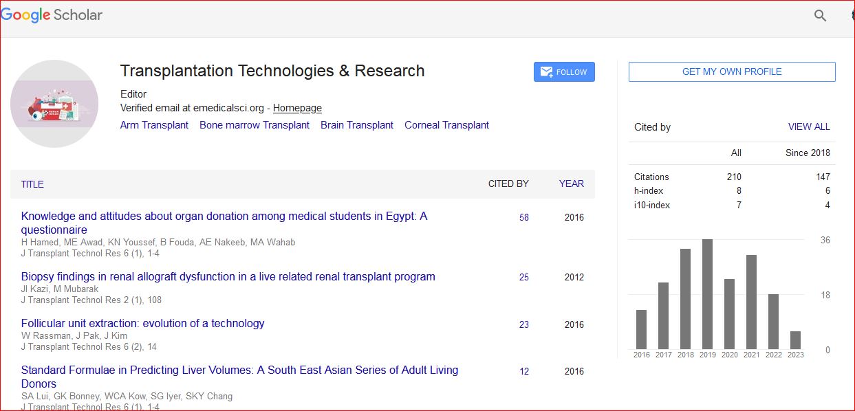
Transplantation Technologies & Research received 223 citations as per Google Scholar report