Opinion - (2022) Volume 7, Issue 4
Received: 04-Jul-2022, Manuscript No. jib-22-77632;
Editor assigned: 06-Jul-2022, Pre QC No. P-77632;
Reviewed: 20-Jul-2022, QC No. Q-77632;
Revised: 25-Jul-2022, Manuscript No. R-77632;
Published:
31-Jul-2022
, DOI: 10.37421/2476-1966.2022.7.182
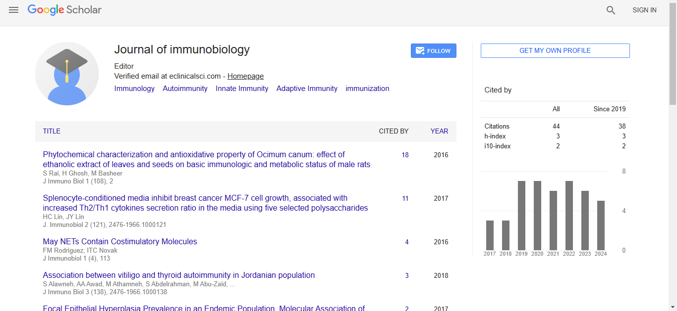
Citation: Postow, Michael. “Immunotoxicity from Chemotherapy Using Checkpoint Inhibitors” J Immuno Biol 7 (2022): 182.
Copyright: © 2022 Postow M. This is an open-access article distributed under the terms of the Creative Commons Attribution License, which permits unrestricted use, distribution, and reproduction in any medium, provided the original author and source are credited.
A growing number of cancers, such as non-small-cell lung cancer (NSCLC), melanoma, urothelial cancer, head and neck, and renal cell cancers, are treatable with anti-cancer immunotherapy. Administration of monoclonal antibodies (mAb) to regulatory immunological checkpoint molecules, which prevent T-cell activation, is currently the most used method. Immune checkpoint inhibitors (ICI) are predicted to be used more often as new therapeutic indications are investigated in clinical studies.
Immune checkpoint inhibitors' primary mechanism of action, immune system dysregulation, is characterized by an overlapping series of immunerelated adverse events (irAEs). As patterns of toxicity differ from those brought on by cytotoxic chemotherapy or molecularly targeted medicines, management of the situation necessitates significant care. To prevent the needless morbidity and mortality linked to the more severe forms of toxicity, it is essential to be aware of and early recognize irAEs. Adverse effects from immunotherapy, in contrast to other systemic anti-cancer therapies, can happen months to years after the final dose, a discovery that has led to the continuation of patient monitoring beyond the end of the therapy.The most prevalent or clinically important toxicities that impact the lungs, gut, liver, and endocrine system are the main emphasis of this article. Published guidelines provide thorough clinical advice on toxicity impacting other systems [1].
Respiratory toxicity
While other clinicopathological entities including sarcoid-like reactions and pleural effusions are infrequent, diffuse inflammation of the lung parenchyma (pneumonitis) is the most common manifestation of ICI-induced pulmonary damage. In clinical studies, pneumonitis has been reported in 2-5% of patients using anti-PD-1 monotherapy; however, the frequency in the real world may be higher (19%). It results in death in 1%–2% of patients, making it the most clinically serious ICI-associated toxicity.
Although this may be explained by the higher number of PD-1/programmed cell death protein ligand 1 (PD-L1) inhibitors in NSCLC trials, the association of pneumonitis with PD-1 axis inhibition is stronger than with that of cytotoxic T-lymphocyte antigen 4 (CTLA-4), and can be enhanced by combined therapy. Pneumonitis rates have been reported to be lower with PD-L1 inhibitors than with PD-1 inhibition in various studies. NSCLC and RCC both have higher rates of pneumonitis than melanoma, with NSCLC having a more rapid onset (2 months) and more severe form, which may be attributable to prior radiotherapy and pulmonary co-morbidities [2].
The median onset time is 3 months, with a range of 1 to 2 years. The earliest signs of dry cough, dyspnea, and hypoxia are non-specific and might be mistaken for other conditions such infection, pulmonary embolism, disease progression or pseudo-progression, and worsening of co-existing lung conditions.
High-resolution computed tomography is the main investigation when it comes to pneumonitis. Non-specific interstitial pneumonia, organizing pneumonia, and hypersensitivity pneumonitis are among the radiographic abnormalities that frequently affect the lower lobes. When there is a clinical suspicion of infection or a lack of response to immunosuppression, bronchoalveolar lavage helps in diagnosis [3].
A CD4+-predominant lymphocyte population, organized pneumonia, diffuse alveolar injury, and granulomatous inflammation are among the histological features of pneumonitis. Although some of these results are similar to those of lung fibrosis in its early stages, pneumonitis typically has a good prognosis. Findings linking lung fibrosis to PD-1 axis activity may help to explain this, at least in the anti-PD-1 context. Anti-PD-1 antibody therapy in mice with idiopathic pulmonary fibrosis reduces fibrosis, but injection of PD-L1- overexpressing fibroblasts into animals increases fibrosis.
According to the severity of the condition, corticosteroids, ICI stoppage, and supportive treatments are used to treat pneumotoxicity. Grade 1 pneumonitis, which accounts for one-third of cases with subclinical radiographic alterations, is managed with treatment withdrawal until spontaneous remission. Mild dyspnea and cough are the hallmarks of grade 2 pneumonitis, which can be treated with oral corticosteroids that are reduced after the onset of symptom relief for at least a month. Hospitalization is indicated by grades 3 and 4 (20%–40% of patients). They are distinguished by severe or life-threatening symptoms, and high-dose intravenous corticosteroids are beneficial in both cases [4].
Intestinal toxicity
Among the most frequent irAEs are toxicities of the gastrointestinal (GI) tract. These can be very serious; 11% of ipilimumab-related colitis deaths have been reported and about one-third of ICI toxicity deaths are attributed to gastrointestinal issues.
The beginning of these GI side effects can occur at any time after treatment has begun, with anti-PD-1 side effects typically manifesting later than anti- CTLA-4 side effects36. However, there have been a few uncommon instances of GI toxicity manifesting up to a year after medication has been stopped. Although clinically severe colitis is the most feared consequence, diarrhea is the most typical GI toxin presentation. Abdominal pain, hemotochezia, fever, and other GI or constitutional symptoms can all be signs of colitis [5].
Unknown mechanisms underlie the GI toxicity caused by ICI treatment. It has been shown that the majority of individuals with inherited mutations in the CTLA-4 gene locus experience gastrointestinal disturbances (diarrhea, enteropathy). This effect may be explained by the loss of CTLA-4 expression in regulatory T cells, which affects peripheral physiological immunosuppression. The significance of regulatory T cells in disease pathogenesis and the crucial function of CTLA-4 therein are highlighted by animal models of inflammatory bowel disease (IBD). Additionally, research on human genetics has revealed a link between IBD and polymorphisms in the CTLA-4 and PD-1 genes. Given the clinical and pathological similarities, it is not surprising that ICI colitis is being treated with a variety of IBD treatments [6].
Liver toxicity
One of the most typical irAEs from ICI is liver damage. Elevations in either aspartate or alanine transaminase are indicators of hepatotoxicity, which is typically asymptomatic. The most severe groups of patients can be riskstratified by using indicators of synthetic malfunction, such as bilirubin and clotting indices. Grade 3–4 damage occurs in 1%–2% of patients receiving single-agent immunotherapy, and hepatitis will develop in 5%–10% of individuals. Hepatitis is more common in dual therapy, occurring in 25%–30% of patients in any grade and up to 15% in grades 3-4.
The lung and endocrine pancreas are two organs that are preferentially impacted by anti-PD-1 ICI, whereas the pituitary and gut are more impacted by CTLA-4 axis suppression. There have been a variety of histopathology findings, and parallels between ICI-associated toxicity and other autoimmune illnesses have been made; a picture peculiar to ICI has emerged in the liver but not the lung. Of course, regulatory T-cell malfunction has been a recurring pattern in all of the tissues examined. Possible complement activation in the pituitary caused by anti-CTLA-4 antibody is one noteworthy mechanism. Guidelines for the management of irAEs have been produced by a variety of scientific associations. These guidelines are based on stopping the medication, using steroids and further immunosuppression, as well as taking other symptomatic treatments. Recognition and quick treatment of irAEs are crucial to reduce patient morbidity given that immunotherapy is presently being tested in more cancer types in addition to the numerous tumour types where it is already commonly employed. To better understand the complicated pathophysiology that underlies the toxicity and responsiveness to ICI in cancer patients, translational studies that provide a more thorough clinical and biological phenotyping of ICI recipients are necessary.
Google Scholar, Crossref, Indexed at
Google Scholar, Crossref, Indexed at
Google Scholar, Crossref, Indexed at
Google Scholar, Crossref, Indexed at
Google Scholar, Crossref, Indexed at
Journal of Immunobiology received 34 citations as per Google Scholar report