Editorial - (2022) Volume 8, Issue 2
Received: 21-Feb-2022, Manuscript No. JCTT-22-55113;
Editor assigned: 24-Feb-2022, Pre QC No. P-55113;
Reviewed: 27-Feb-2022, QC No. Q-55113;
Revised: 28-Feb-2022, Manuscript No. R-55113;
Published:
11-Mar-2022
, DOI: 10.4172/2471-9323.22.8.176
Citation: Varthya, Shoban Babu. “Overview of Skin Conditions.” J Cosmo Tricho 8 (2022): 176. DOI: 10.4172/2471-9323.22.8.176
Copyright: © 2022 Varthya SB. This is an open-access article distributed under the terms of the Creative Commons Attribution License, which permits unrestricted use, distribution, and reproduction in any medium, provided the original author and source are credited.
The human integumentary system, which is made up of skin, hair, nails and associated muscle and glands and covers the whole surface of the body, is affected by a variety of disorders. The epidermis, dermis and subcutaneous tissue make up the epidermis, which weighs an average of four kilos and covers an area of two square metres. Glabrous skin, which is hairless on the palms and soles and hair-bearing skin are the two basic varieties of human skin. Hairs in the latter type grow in structures called pilosebaceous units, which contain a hair follicle, a sebaceous gland and a muscle called the arrector pili. The epidermis, hair and glands are formed by the ectoderm in the embryo, which is impacted chemically by the underlying mesoderm, which develops the dermis and subcutaneous tissues.
The epidermis is the skin's most superficial layer, consisting of numerous strata: stratum corneum, stratum lucidum, stratum granulosum, stratum spinosum and stratum basale. Because the epidermis lacks a direct blood supply, nutrients are delivered to these layers via diffusion from the dermis. Keratinocytes, melanocytes, Langerhans cells and Merkel cells are the four cell types that make up the epidermis. Keratinocytes are the most common kind, accounting for around 95% of the epidermis. Cell division within the stratum basale, in which developing cells steadily shift outwards via the stratum spinosum to the stratum corneum, where cells are constantly shed from the surface, keeps this stratified squamous epithelium alive. In healthy skin, production equals loss; it takes about two weeks for a cell to migrate from the basal cell layer to the top of the granular cell layer and requires next 2 weeks to pass the stratum corneum [1-3].
The dermis is the layer of skin that lies between the epidermis and the subcutaneous tissue and it is divided into two sections: papillary and reticular. The overlaying rete ridges of the epidermis interdigitate with the superficial papillary dermis and the two layers interact through the basement membrane zone. Collagen, elastic fibres and ground material are structural components of the dermis. The pilosebaceous units, arrector pili muscles and eccrine and apocrine glands are among these components. The dermis has two circulatory networks, one superficial and one deep plexus that run parallel to the skin surface and are joined by vertical communication veins. Blood vessels in the dermis have four functions: they deliver nutrients, regulate temperature, moderate inflammation and help in wound healing [4,5].
The subcutaneous tissue is a fat layer that lies between the dermis and the fascia underneath it. The real fatty layer, or panniculus adiposus and a deeper vestigial layer of muscle, the panniculus carnosus, can be separated from this tissue. The adipocyte, or fat cell, is the major biological component of this tissue. This tissue is divided into septal and lobular compartments, which have different microscopic appearances. Subcutaneous fat functions as a reserve energy source, insulates the body, absorbs shock and insulates the body. The human integumentary system includes a wide spectrum of illnesses, known as dermatoses, as well as a number of non-pathologic ailments.
The author shows no conflict of interest towards this manuscript.
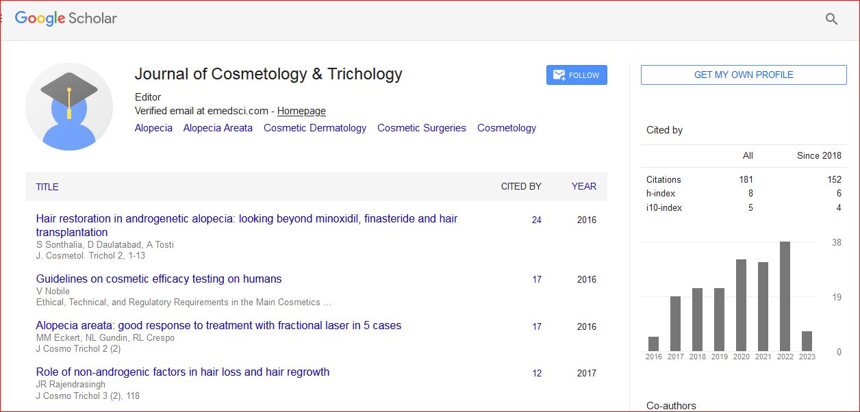
Journal of Cosmetology & Trichology received 180 citations as per Google Scholar report