Research Article - (2024) Volume 10, Issue 1
Received: 14-Dec-2023, Manuscript No. jpnp-23-122830;
Editor assigned: 16-Dec-2023, Pre QC No. P-122830;
Reviewed: 15-Jan-2024, QC No. Q-122830;
Revised: 24-Jan-2024, Manuscript No. R-122830;
Published:
02-Feb-2024
, DOI: 10.37421/2472-0992.2024.10.281
Citation: Tumanjong, Irene Memeh, Tobias Obejum Apinjoh,
Faustin Pascal Tsague Manfo and Evans Ngandung Mainsah, et al. “In Vitro Antionchocercal Activity, Phytochemical Analysis and Toxicity Studies of Extracts of Azadirachta indica.” J Pharmacogn Nat Prod 10 (2024): 281.
Copyright: © 2024 Tumanjong IM, et al. This is an open-access article distributed under the terms of the Creative Commons Attribution License, which permits unrestricted use, distribution, and reproduction in any medium, provided the original author and source are credited.
Ivermectin and moxidectin are the only recommended drugs for the treatment of onchocerciasis, with the former being the most widely used. However, both drugs are not suitable in eliminating the disease. There is the need to identify novel anti-onchocercal agents including from plant sources. This project investigated the anti-onchocercal activity of extracts of Azadirachta indica that could eventually yield new drug leads for the cure of onchocerciasis. Organic extracts were obtained from the leaves and seeds of Azadirachta indica using solvents of different polarities and tested in vitro against two developmental stages of the bovine model parasite, Onchocerca ochengi. Both microfilariae (mf) and adult male worm viabilities were assessed by motility reduction, while adult female worm viability was evaluated using the standard MTT/formazan assay. Toxicity of active extracts was assessed on monkey kidney epithelial cells (LLC-MK2) and in BALB/c mice. The methylene chloride extract of the leaves was the most active against the adult female worms and the mf with IC50s of 55.61 μg/ml and 8.048 μg/ml respectively. The hexane extract of the leaves was the most active against the adult male worms with an IC50 of 16.34 μg/ml. Selectivity indices for the most active extracts were 1.12 for adult females, 7.77 for the mf and 7.35 for adult males indicating that the extracts are selectively active on the parasites. The most active extracts showed no acute toxicity to Balb/c mice and had no significant effect on the liver enzymes, alanine aminotransferase and aspartate aminotransferase and markers of kidney function, urea and creatinine (p<0.05). Phytochemical analysis revealed the presence of saponins, flavonoids, steroids, tannins, alkaloids, polyphenols and terpenoids. The anti-onchocercal activity and selectivity indices of A. indica extracts suggest the plant is a potential source of new anti-onchocercal drug leads justifying further investigations for the identification and isolation of the bioactive compounds.
Onchocerciasis • Ivermectin • Onchocerca ochengi • Azadirachta indica • Toxicity • Phytochemical analysis
Human onchocerciasis, also known as ‘river blindness’ is a chronic skin and eye disease that afflicts millions of people residing in many fertile riverine areas of tropical countries (mostly in sub Saharan Africa) [1]. Currently, there is an estimated 187 million people at risk of getting sick with onchocerciasis and it is also estimated that some 37 million people are infected with O. volvulus. Among those infected, an estimated 6.5 million live with skin manifestations of the disease and 2 million are estimated to be either visually impaired or blind. About 99% of all reported cases of the disease are living in 31 endemic countries in Sub-saharan Africa with a small percentage living in Yemen and latin America [2] Onchocerciasis causes chronic suffering and severe disability with approximately 1.5 million disability-adjusted life-years (DALYs) being lost each year due to this disease [3]. Cameroon has a high prevalence of the disease; with studies reporting a prevalence of 40% in districts in the West region [4]. It is the second leading infectious cause of blindness globally and it is caused by infection with O. volvulus, a parasitic worm transmitted through the bites of blood feeding blackflies (Simulium spp) [2]. Some clinical symptoms associated with the disease can include intense itching, eye disease, epilepsy, as well as disfiguring skin lesions caused by the microfilariae (mf), which move around the human body in the subcutaneous tissue and induce intense inflammatory responses when they die [5,6]. The burden of the disease results in abandonment of fertile farmlands which affects the poorest rural communities in Africa often in areas where subsistence farming is of vital economic importance, as well as long term disability and social stigmatization [7,8].
Global efforts put in place to eliminate onchocerciasis have been based largely on chemotherapy, which is by Mass Drug Administration (MDA) through annual Community Directed Treatment with Ivermectin (CDTI) [9]. However, ivermectin is only microfilaricidal and requires annual treatment with a certain degree of therapeutic coverage and which typically lasts at least 12–15 years, corresponding to the reproductive lifespan of the adult worm [10]. Moreover, ivermectin cannot be safely used in areas where Loa loa is prevalent as treatment of onchocerciasis patients who are co-infected with L. loa mf has been associated with serious adverse events, including encephalopathy and death [11-13]. Lastly, resistance to ivermectin has been reported in some parasitic nematodes in veterinary medicine and it is thought that it may extend to the human O. volvulus [14,15]. Moxidectin, which was recently approved is indicated for the treatment of onchocerciasis at a single oral 8 mg dose for individuals ≥ 12 years [16,17]. Moxidectin is however only microfilaricidal and the safety and efficacy of repeated administration in patients with Onchocerca volvulus have not been studied. The use of both drugs does not meet the goal of achieving elimination of human onchocerciasis in the foreseeable future. There is therefore the need for alternative anti-onchocercal agents.
Medicinal plant preparations have for a long time been identified as alternative remedies for several diseases [18] and also for the identification of potential drug leads. Many drugs used in modern medicine today are of plant origin and most of Africa’s population rely on medicinal plants for their health needs [19]. Some medicinal plants have been shown to have antionchocercal effects [20] and there is a continuous search for more of such medicinal plants. Many of the studies have made use of the bovine model, O. ochengi, which is currently known to be the closest relative and most suitable model of O. volvulus, because of serious difficulties involved in obtaining O. volvulus from humans [21]. In this regard, Azadirachta indica, which is commonly known as neem is well known for its medicinal use. It belongs to the family Meliaceae and is widely distributed within tropical regions especially in India and West Africa [22]. Almost every part of this tree is a good source of bioactive compounds and some of the phytochemicals present in this plant have been isolated, quantified and identified through intensive studies. Previous chemical analysis of the plant has identified various phytoconstituents such as alkaloids, flavonoids, terpenoids, phenols, reducing sugars, glycosides, tannins, cyclic trisulphide, oxalic acid, steroids, fatty acids and saponins [23-25] with several pharmacological effects such as antimicrobial, antifungal, antipyretic, antiproliferative, anti-cancer and anti-parasitic properties [24,26-29]. Some studies have demonstrated the anthelminthic activity of A. indica, such as that of Jamra N, et al. [30] who evaluated the anthelmintic efficacy of crude A. indica leaf powder against strongyle infections in cattle. Another study is that of Sharba K [31] who investigated the in vitro anthelmintic activity of A. indica leaves on the microfilariae of Setaria cervi. The plant has been shown to be very effective in the removal of intestinal worms [26]. Also, a study by Mishra V, et al. [32] demonstrated the antifilarial activity of alcohol and aqueous extracts of flowers of A. indica in vitro against the mf of S. cervi. Ethnobotanical surveys carried out among traditional healers in the Far-North region of Cameroon by Mamat A, et al. [33] showed that A. indica is amongst the medicinal plants used in the management of onchocerciasis. However, there is no available scientific data backing the use of this plant in the treatment of onchocerciasis.
This study was therefore aimed at investigating the in vitro antionchocercal activity of crude extracts of A. indica as a starting point, against the bovine model parasite, O. ochengi. The cytotoxicity and acute toxicity of active extracts were also investigated. This could serve as a potential source of new lead compounds for the development of safe and efficacious drugs against onchocerciasis.
Collection, identification and processing of plants
A. indica plant parts were collected based on ethno pharmacological information from Maroua in the Far North Region of Cameroon. Voucher specimens were taken to the Yaounde´ herbarium where they were authenticated by Eric Ngansop and voucher number was assigned to it (A. indica: 4447/SRFK). The leaves and seeds of the plant were air dried to constant weight and then ground to fine powder using a grinding mill. Each powder was macerated for 48 hours, sequentially, in hexane, methylene chloride and methanol, with the collection of filtrate after the use of each solvent. The filtrates were concentrated using a rotary evaporator (BUCHI Rotavapor R-200, Switzerland) at the boiling points of each solvent. Each crude extract was recovered and left on the shelf for 72 hours for any residual solvent to evaporate.
The crude extracts were then weighed and the percentage yield in extract was calculated using the following formula:
% yield = (Weight of crude extract × 100) /Weight of dry grounded plant material
The crude extracts were preserved at -20 ℃ for later use. A stock solution of 25 mg/mL was prepared in >99.8% DMSO (Sigma, Germany) and further diluted in culture medium for assays on the different worm stages.
Isolation of O. ochengi adult worms
Adult worms were isolated from umbilical cattle skin as previously described by Cho-Ngwa F, et al. [34]. The plates containing the worms were incubated overnight at 37 ℃ under an atmosphere of 5% CO2 in humidified air in a HERACELL-150i CO2 incubator (USA). Prior to drug testing in the primary and secondary screens, the viability of worms and sterility of cultures were evaluated by visual and microscopic examination using an inverted microscope (Nikon Eclipse TS100, China).
Mammalian cells for microfilarial cultures and cytotoxicity studies
Monkey kidney epithelial cells (LLC-MK2) obtained from the American Type Culture Collection (ATCC, USA), were proliferated in CCM at 37°C under an atmosphere of 5% CO2 in humified air until they became fully confluent. Once the cells became fully confluent, the old media was decanted and the cells were dislodged with 0.125% trypsin and 0.5 mM EDTA in serum-free RPMI-1640 culture medium. The dislodged cells were re-suspended in 10 mL of complete culture medium and centrifuged at 560 × g for 10 minutes to get rid of the trypsin. The last procedure was repeated once. The cell suspension (100 μL/well) was transferred into 96-well microtitre culture plates and kept in the CO2 incubator for cells to grow and become fully confluent, usually taking 3-5 days depending on the initial concentration of cells. These cells served as feeder layer for O. ochengi microfilariae assays and also for cytotoxicity studies.
Isolation and culturing of O. ochengi microfilariae
Isolation of O. ochengi microfilariae (mf) was done as previously described [34] with slight modifications. Cattle skin with palpable nodules obtained from slaughter houses in Douala and Buea, Cameroon, carefully washed and dabbed. A few (about 5) skin snips were obtained from different locations of the skin and incubated in small amounts of culture medium for 30 minutes. Emerged microfilariae were qualified and quantified with the aid of an inverted microscope. Only pieces of skin containing large amounts of microfilariae of which >90% were O. ochengi were selected for the assay. The selected skin was sterilized as above, tautly attached onto a sterile wooden board using autoclaved thumbnails and was then carefully shaved with a sterile razor blade, after which it was washed with distilled water and sterilized with 70% ethanol. Skin slivers (about 0.5 cm wide) of epidermis and dermis were obtained and incubated for 4-6 hours at room temperature in complete culture medium (CCM) in 50 ml Falcon tubes. The emerged and highly motile O. ochengi mf were concentrated by centrifugation at 400 × g for 10 minutes and quantified microscopically. The mf were re-suspended in CCM and distributed into 96- well microtitre culture plates containing LLC-MK2 cell layer, with each well containing 10-15 mf in 100 μl of CCM. Prior to addition of drugs, the viability of the worms and sterility of cultures were ascertained by microscopy.
Primary screens on O. ochengi adult worms
All extracts were screened on O. ochengi adult worms. The worms were treated in triplicates with an extract at 500 μg/ml in CCM, with auranofin at 10 μM, serving as positive control [35] and 2% DMSO as negative control. The treated worm cultures were incubated for 5 and 7 days for male and female worms, respectively, at 37°C in 5% CO2 atmosphere and their viability assessed. Activity against adult female worms was assessed using the standard MTT/Formazan assay for testing biochemical death. Activity scores assigned ranged from 100% parasite killing (no blue formazan coloration seen), through 90%, 75%, 50%, 25%, to 0% (entire worm appears blue as in negative control). For the male worms, motility scoring based on microscopy was used. Activity scores ranged from 100% (complete inhibition of motility), 75% (only head or tail of worm shaking occasionally), 50% (whole worm motile, but sluggishly), 25% (only little change in motility), to 0% (no observable change in motility as in negative control).
An extract was considered active if there was ≥ 90% inhibition of male worm motility or of formazan formation; moderately active if there was a 50– 89% inhibition of male worm motility or of formazan formation and inactive if there was a <50% inhibition of male worm motility or of formazan formation. All experiments were repeated at least once before secondary screens could be done.
Secondary screens on O. ochengi adult worms
Extracts with 100% activity on adult worms at the primary screens were re-tested as described under primary screens at serial dilutions of seven concentrations (from 500 to 7.8125 μg/mL), in order to determine their IC50 values.
Primary screens on O. ochengi microfilariae
The assays were conducted at 500 μg/mL in duplicates in 96 well microtitre plates. The mf were incubated with drugs for a period of 5 days, at 37 oC under an atmosphere of 5% CO2 in humidified air in a total volume of 200 μl of CCM medium. The positive control drug was amorcarzine at 10 μM and 2% DMSO served as negative control. Viability of the mf was assessed microscopically, daily, by assigning percentage motility inhibition scores as 100% (all mf immotile), 75% (only head or tail of mf shaking, occasionally), 50% (whole body of mf motile but sluggishly or with difficulties), 25% (almost vigorous motility), 0% (vigorous motility as with negative control). Every screen was repeated at least once.
Secondary screens on O. ochengi microfilariae
Extracts that showed 100% activity on the mf at primary screens were retested as described under the primary screens on mf at serial dilutions of seven concentrations (ranging from 500 to 7.8125 μg/mL), in order to determine the IC50 values.
Toxicity assessment
Cytotoxicity studies: Cytotoxicity of interesting extracts was assessed on LLC-MK2 cells on day 5 as part of the mf secondary screen assays. The 50% cytotoxic concentrations (CC50 values) for these cells were determined by microscopic examination. Dead or deformed cells were usually detached from the bottom of the vessel and were rounded in shape. The morphological deformation data was used to estimate the CC50 values for these cells. The Selectivity Index (SI) of each extract was calculated as the ratio of CC50 of the extract on the mammalian cells to the IC50 of the extract on the O. ochengi parasite.
Acute toxicity assessment: This assay was conducted in accordance with the Organisation for Economic Co-operation and Development (OECD) guidelines for testing chemicals [36]. Ethical approval (2019/018/UB/IACUC/ BTU/FS) for the use of the animals was obtained from the Institutional Animal Care and Use Committee (UB-IACUC), University of Buea, Cameroon. Acute toxicity was evaluated for the most active extracts on O. ochengi adult worms and mf. Briefly, 18 BALB/c mice comprising one control and two test groups of six animals each (10 weeks old, three females and three males per group) from the animal house of the Biotechnology unit were selected and kept separately in their respective cages for one week, to allow for acclimatization to the new housing conditions. Each of the two treatment groups received one of the extracts at a limit dose of 2000 mg/kg body weight, administered orally with a volume of 1 mL/100 g body weight of mice, while the control group received the diluent only (2% DMSO). The animals were observed for any gross changes in physical activity and behavioural pattern, food and water intake, stool sample, loss of fur, sensitivity to sound, sensitivity to pain, sleep pattern, coma and death every day for 14 days. The animals were then fasted overnight and anaesthetized with ketamine/xylazine (90/10 mg/kg) and blood was collected by retroorbital bleeding. The blood was coagulated for 30 minutes and centrifuged at 2200 rpm for 15 minutes to obtain serum which was then used to measure the activity of liver enzymes, alanine aminotransferase and aspartate aminotransferase and also to measure markers of kidney function, urea and creatinine, using Chronolab Systems Diagnostic Kits (Barcelona, Spain), following the manufacturer’s instructions.
Qualitative phytochemical screening
The presence of the major groups of phytochemicals in the most active extracts were investigated using standard procedures: Liebermann-Buchard test for steroids, salkowski test for terpenoids, FeCl3 test for polyphenols, Stankovic test for flavonoids, Keller-killiani test for cardiac glycosides, Ferric chloride test for tannins, Wagner’s test for alkaloids and frothing test for saponins [37].
Statistical analyses
The data obtained were analysed using GraphPad Prism 8.0 (GraphPad Prism INC., CA, USA) to obtain IC50 values of active extracts. The logarithm of the extract concentration was plotted against its activity determined by microscopy, to obtain a nonlinear regression curve –fitting and a variable slope sigmoidal dose- response curve. CC50 values were derived from a plot of % inhibition of cell viability against log of extract concentration generated using the same software. The same software was used to analyse liver enzymes and markers of kidney function. An unpaired two-tailed t-test was used to check for any significant difference (at P<0.05) between the control and test groups.
A total of 6 extracts were obtained from the leaves and seeds of the plant, A. indica using solvents of different polarities in a sequential order: hexane followed by methylene chloride and then methanol. The hexane extract of the seeds had the highest yield (7.6%) while the methanol extract of the leaves had the lowest yield (1.7%), (Table 1).
| Plant | Plant part | Solvent | Code | %Yield |
|---|---|---|---|---|
| Azadirachta indica | Leaves | Hexane | AILhex | 2.8 |
| Leaves | Methylene chloride | AILmc | 3.8 | |
| Leaves | Methanol | AILmet | 1.7 | |
| Seeds | Hexane | AIShex | 7.6 | |
| Seeds | Methylene chloride | AISmc | 3.5 | |
| Seeds | Methanol | AISmet | 5.6 |
In the primary screens, at 500 μg/ml, 3 out of the 6 extracts showed 100% activity against the adult male worms while 2 showed 100% activity against the adult female worms. In the secondary screens, the IC50 values ranged from 16.34 to 164.4 μg/mL on adult male worms with the hexane extract of the leaves (AILhex) being the most active with an IC50 value of 16.34 μg/mL. The IC50 values of the 2 extracts that showed 100% activity on adult female worms were 55.61 and 151.2 μg/mL with methylene chloride extract being the most active. The extracts showed a dose-dependent activity as illustrated for the most active extracts in Figure 1, with an overall higher activity against adult male worms.

Figure 1. Dose dependent effect of A) AILhex on adult O. ochengi male worms, B) AILmc on adult O. ochengi female worms and C) AILmc on O.ochengi microfilariae.
On the mf, 4 out of the 6 extracts screened at 500 μg/mL in the primary screens showed 100% activity. In the secondary screens, the IC50 values ranged from 8.048 to 44.19 μg/ml; the methylene chloride extract of the leaves (AILmc) was the most active on the mf with an IC50 value of 8.048 μg/ml. A dosedependent activity was also illustrated for the most active extract as shown in (Figure 1).
The cytotoxic effects of extracts with high activity were evaluated on monkey kidney epithelial cells (LLC-MK2) on day 5 of culturing in CCM and as part of the mf assay. The extract concentration inducing cytotoxicity in 50% of cells (CC50) was determined and their Selectivity Indices (SI) calculated. AILhex was more toxic to O. ochengi female worms than to the cells while AILmet was more toxic to O. ochengi male worms than to the cells as reflected by their selectivity indices. On O. ochengi mf, AILhex recorded the highest SI of 7.87 while AISmet recorded the lowest SI of 4.28. On adult males, AILhex recorded the highest SI of 7.35, while AILmet recorded the lowest SI of 0.76. Finally, on adult females, AILmc recorded an SI value of 1.12 as opposed to AILhex which recorded an SI value of 0.79 (Table 2).
| Extract code | CC50 (μg/mL) | IC50 (μg/mL) | Selectivity index (CC50/IC50) | ||||
|---|---|---|---|---|---|---|---|
| Cells | AM | AF | MF | AM | AF | MF | |
| AILhex | 120.1 | 16.34 | 151.2 | 15.26 | 7.35 | 0.79 | 7.87 |
| AILmc | 62.50 | 27.80 | 55.61 | 8.048 | 2.25 | 1.12 | 7.77 |
| AILmet | 125.2 | 164.4 | NR | 27.80 | 0.76 | NR | 4.50 |
| AISmet | 189.2 | NR | NR | 44.19 | NR | NR | 4.28 |
The most active extracts, AILhex and AILmc, on O. ochengi adult male and female worms, respectively, showed no sign of acute toxicity 14 days posttreatment when administered orally at a single limit dose of 2000 mg/kg body weight in Balb/c mice. The average weights of the mice increased from 21.1g pre-treatment to 22.5g post-treatment. When compared with the control group, there was no significant difference in food and water intake. No change in physical appearance, physical activity and behaviour was observed, likewise there was no loss of fur and no change in skin and mucous membranes.
In order to compare the effects of AILmc and AILhex on liver enzymes, an unpaired two-tailed t-test was done for Aspartate Aminotransferase (AST) and Alanine Aminotransferase (ALT) at 95% confidence interval (P<0.05) as both control and test groups had the same variances. There was a significant difference between the control (2% DMSO) and test group treated with AILmc for the enzyme AST with a P value of 0.0200 while there was no significant difference between the control and test group treated with AILmc for the enzyme ALT with a P value of 0.2259 as shown in Figure 2. The AST:ALT ratio of mean enzyme activity which indicates the level of liver injury for the test group was =1.11, indicating a negligible effect in the mouse liver given it is slightly >1 (Figure 2).

Figure 2. Effect of 2000 mg/kg methylene chloride extract of Azadirachta indica (AILmc) on mouse liver enzyme activity. AST: Aspartate Aminotransferase (P=0.0200); ALT: Alanine Aminotransferase (P=0.2259).
There was no significant difference between the control group and mice treated with AILhex for enzyme activities with P values of 0.6069 and 0.5338 for AST and ALT respectively, as shown in Figure 3. The AST:ALT ratio was 1.0 indicating no adverse effect in the mouse liver given it is =1 (Figure 3).

Figure 3. Effect of 2000 mg/kg hexane extract of Azadirachta indica (AILhex) on mouse liver enzyme activity. AST: Aspartate Aminotransferase (P=0.6069); ALT: Alanine Aminotransferase (P=0.5338).
Serum urea and creatinine are good indicators of a normal functioning kidney and an increase in their levels is indicative of kidney dysfunction. In order to assess the effects of AILmc and AILhex on the kidney, urea and creatinine levels were measured in serum samples from mice treated with the extracts. As shown in Figures 4 and 5 there was no significant difference between the control group mice and mice treated with the extracts for urea (with P values of 0.5091 and 0.5154, for AILmc and AILhex, respectively) and creatinine (with P values of 0.231 and 0.4654, for AILmc and AILhex, respectively) (Figures 4 and 5).

Figure 4. Effect of 2000 mg/kg methylene chloride extract of Azadirachta indica (AILmc) on kidney metabolites; Urea (P=0.5091) and creatinine (P =0.2312).

Figure 5. Effect of 2000 mg/kg hexane extract of Azadirachta indica (AILhex) on kidney metabolites; Urea (P=0.5154) and creatinine (P =0. 4654).
Screening of the two most active extracts on adult worms, AILmc and AILhex, revealed the presence of several categories of phytochemicals including saponins, flavonoids, steroids, tannins, alkaloids, polyphenols and terpenoids (Table 3).
| Extract | Saponins | Flavonoid | Steroids | Tannins | Alkaloids | Cardiac glycoside | Polyphenol | Terpenoids |
|---|---|---|---|---|---|---|---|---|
| AILmc | + | ++ | + | + | ++++ | - | + | ++ |
| AILhex | ++ | + | +++ | + | + | - | +++ | +++ |
The aim of this study was to assess the activity of the medicinal plant, A. indica on O. ochengi worm stages as an early step in the search of novel anti-onchocercal drugs. In Cameroon, this plant has traditionally claims in the treatment of several diseases such as; diabetes, HIV/AIDS, cancer, cardiovascular diseases, skin infections, malaria, ulcers and hepatitis. Previous research has suggested that the plant also possesses a number of pharmacological properties. These include anticancer, immunomodulatory, antidiabetic, neuroprotective, anti-inflammatory, antiviral, antibacterial, antifungal, antioxidant, antimalarial and wound healing activities [38]. A. indica was selected based on previous pharmacological properties demonstrated on other helmintic diseases and the anti-onchocercal potential of the extracts of its leaves and seeds were determined. A total of 6 crude extracts of varying solubilities were prepared from the leaves and the seeds of the plant and tested in already standardised assays. From the results obtained, there was a general decrease in adult worm and mf viabilities with increasing concentration of active extracts (Figure 1). This indicates the typical dose dependency characteristic of substances used as drugs. A time dependency characteristic was also demonstrated as confirmed by the fact that the anti-parasitic activities of the extracts increased with increase in time of incubation in drug containing medium.
The methylene chloride and hexane extracts of A. indica leaves investigated in the present study exhibited promising anti-onchocercal activity on the different worm stages of O. ochengi and also recorded minimal cytotoxic effects. The methylene chloride extract was most active on the adult female worms and mf while the hexane extract was most active on the adult male worms. Solvent extracts of the leaves have shown significant anthelmintic activity against the microfilariae of S. cervi with the highest mortality rate observed in methanol and ethanol extracts [31], as opposed to methylene chloride extract of the leaves, the latter rather recorded the highest mortality rate against O. ochengi mf in our study (Figure 1). This difference might be due to differences in worm species and in the proteins being expressed. Also, this difference could be due to differences in polarity of the extracts as previous studies show non-polar extracts to be more active against the nematode O. ochengi than polar extracts [39-41]. The anthelmintic effect of A. indica leaves extract has been shown on L3 larvae of Haemonchus contortus from goats and it showed 40% mortality of the larvae at 4 mg/ml after 24 h treatment [42]. In another study, the in vitro anthelmintic effect of ethanol extract of A. indica was investigated against gastro-intestinal nematodes of fowl and findings showed 100% efficacy at a concentration of 50 mg/ml [43]. This goes a long way to prove that this plant is a potent anthelmintic agent; however, this study is reporting its activity on the nematode O. ochengi for the first time.
Overall, extracts from the leaves showed higher activity on all the worm stages than extracts from the seeds. The leave extracts had higher activity on the adult male worms (lower IC50s) than on the adult female worms. This difference may be due to the differences in proteins being expressed at the different sexes or in the viability tests used. Generally, all extracts demonstrated higher activity on the microfilariae than on the adult worms, as indicated by their lower IC50s. This might be due to the fact that the mf is smaller than the adult worms or differences in metabolism or in the assays used [39]. Ivermectin, the drug of choice in the control of onchocerciasis is known to exert a similar selective effect. Our findings are in line with the results of Cho-Ngwa F, et al. [44] and Tiku E, et al. [45] whose extracts showed higher activity on the mf than on the adult worms. The promising properties of A. indica warrants further investigation which may lead to the isolation and studies on the mechanisms of action of pure compounds responsible for specific killing of the different O. ochengi worm stages.
It was necessary to generate preliminary safety data on these extracts after investigating their anti-onchocercal activity. This was done by evaluating cytotoxicity on LLC-MK2 cells for 4 extracts that showed activity on any of the worm stages and acute toxicity in Balb/c mice for the 2 most active extracts on the adult worm stage. The hexane extract of the leaves showed high selectivity for the adult male as opposed to the adult female. The methylene chloride extract of the leaves had low selectivity for adult females and the methanol extract of the leaves showed low selectivity for adult males as reflected by their SI values (Table 2). All extracts that showed activity on the mf were relatively safer as reflected by their SI values (Table 2). Low SI values signify that the extracts have the potentials of being toxic to parasite at a rate almost equal to the toxicity to mammalian cells. These low SI values can be compared to that of the control drug; ivermectin which was 1 μg/ml and the drug is still in use till date which suggests that cytotoxicity of an extract/compound might not necessarily imply in vivo toxicity. Also, this cytotoxicity is only indicative and may not predict human toxicity because of the difference in susceptibility of cells from different species to drugs [46] and because of a possible complex nature of the extracts whereby therapeutic and toxic compounds may co-exist therein.
When tested for toxicity in vivo, the 2 most active extracts on the adult worm (AILmc and AILhex) showed no sign of acute toxicity to Balb/c mice. This suggests that these 2 extracts could be safely used in phytomedicines for onchocerciasis treatment and justifies their popular use as a natural remedy to many ailments in different parts of the world. It is however better for such usage to be standardized always.
Looking at the effect of these extracts on the liver, there was no significant difference in activities of liver enzymes between the control and test groups except for the enzyme AST in which there was a significant difference between the control and the test group for the extract, AILmc (Figures 2 and 3). The AST:ALT ratio of 1.11 for mice treated with methylene chloride extract was slightly greater than 1 indicating a mild toxicity effect to the liver which points to a mildly reversible injury which may be aggravated at higher doses. Also, the AST: ALT ratio of 1 for the hexane extract indicates absence of toxicity to the liver [47]. This can be compared to the study by Kang TR, et al. [48] which reported an AST:ALT ratio of 0.54 for mice treated with hexane extract of Usnea articulata indicating the absence of toxicity to the liver.
Creatinine and urea are classic markers of kidney function. Their measurement allows for the detection of early metabolic changes resulting from kidney damage [49]. Looking at the effect of the extracts AILhex and AILmc on the kidneys, the differences in serum urea and creatinine levels determined for the mice treated and control groups were not statistically significant indicating that these extracts had no adverse effects on the kidneys.
Qualitative phytochemical analysis of AILhex and AILmc revealed the presence of secondary metabolites; saponins, flavonoids, steroids, tannins, alkaloids, polyphenols and terpenoids (Table 3) in both extracts of the plant. This suggests that although varied, the active principles in the extracts may be from one or more of the aforementioned groups of compounds. These phytochemicals have also been reported in aqueous and ethanolic extracts of this plant [23]. Previous findings have shown such secondary metabolites to be responsible for the medicinal properties of many plants [37,40].
The methylene chloride and hexane extracts of A. indica leaves had significant anti-onchocercal effects on the different worm stages of O. ochengi in a dose-dependent manner with the methylene chloride extract being most active on the adult female worms and mf, while the hexane extract was most active on the adult male worms. The anti-onchocercal activity and minimal toxic effects of extracts of A. indica warrants the use of this plant as a potential source of new drug leads which could be further developed into a cure for onchocerciasis.
All relevant data are within the manuscript. Any other data are available from the corresponding author upon request.
The authors declare that there is no conflict of interest regarding the publication of this article.
Fidelis Cho-Ngwa conceptualized the study, supervised the experiments and participated in the manuscript write-up. Apinjo O. Tobias and Manfo T. F. Pascal co-supervised the study, curated the data and participated in manuscript drafting. Irene Memeh Tumanjong conducted the experiments and participated in manuscript drafting. Evans Mainsah and Gamua Stanley collected the resources and participated in manuscript editing. All authors read and approved the final manuscript.
The authors acknowledge Mr Dieudonne Esoh Achendong of Guidiguis health district, Far-North region of Cameroon, for his assistance in harvesting and transportation of the plant from Maroua, Far-North Region, Cameroon. We will also like to acknowledge the Biotechnology Unit of the Faculty of Science, University of Buea, Buea, Cameroon in general for technical and material assistance. This work was carried out in the Laboratory for Drugs and Molecular Diagnostics Research (ANDI Centre of Excellence for Onchocerciasis Drug Research), Biotechnology Unit, University of Buea, as part of research activities of the authors.
Google Scholar, Crossref, Indexed at
Google Scholar, Crossref, Indexed at
Google Scholar, Crossref, Indexed at
Google Scholar, Crossref, Indexed at
Google Scholar, Crossref, Indexed at
Google Scholar, Crossref, Indexed at
Google Scholar, Crossref, Indexed at
Google Scholar, Crossref, Indexed at
Google Scholar, Crossref, Indexed at
Google Scholar, Crossref, Indexed at
Google Scholar, Crossref, Indexed at
Google Scholar, Crossref, Indexed at
Google Scholar, Crossref, Indexed at
Google Scholar, Crossref, Indexed at
Google Scholar, Crossref, Indexed at
Google Scholar, Crossref, Indexed at
Google Scholar, Crossref, Indexed at
Google Scholar, Crossref, Indexed at
Google Scholar, Crossref, Indexed at
Google Scholar, Crossref, Indexed at
Google Scholar, Crossref, Indexed at
Google Scholar, Crossref, Indexed at
Google Scholar, Crossref, Indexed at
Google Scholar, Crossref, Indexed at
Google Scholar, Crossref, Indexed at
Google Scholar, Crossref, Indexed at
Google Scholar, Crossref, Indexed at
Google Scholar, Crossref, Indexed at
Google Scholar, Crossref, Indexed at
Google Scholar, Crossref, Indexed at
Google Scholar, Crossref, Indexed at
Google Scholar, Crossref, Indexed at
Google Scholar, Crossref, Indexed at
Google Scholar, Crossref, Indexed at
Google Scholar, Crossref, Indexed at
Google Scholar, Crossref, Indexed at
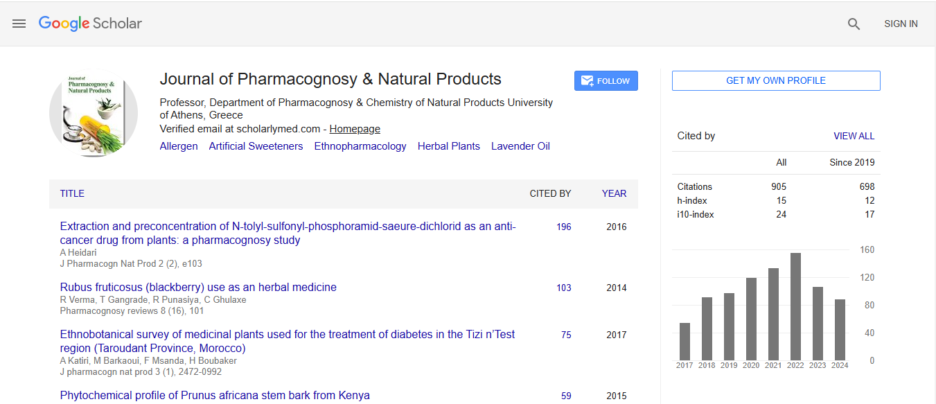
Journal of Pharmacognosy & Natural Products received 606 citations as per Google Scholar report