Commentary - (2025) Volume 15, Issue 3
Received: 13-Jun-2025, Manuscript No. JBL-25- 166574 ;
Editor assigned: 16-Jun-2025, Pre QC No. JBL-25- 166574 (PQ);
Reviewed: 30-Jun-2025, QC No. JBL-25- 166574 ;
Revised: 07-Aug-2025, Manuscript No. JBL-25- 166574 (R);
Published:
16-Aug-2025
, DOI: 10.37421/2165-7831.2025.15.339
Citation: Miki, Hirokazu. ''Hyperthermia against Plasma Cell Dyscrasias" J Blood Lymph 15(2025):339.
Copyright: © 2025 Miki H. This is an open-access article distributed under the terms of the creative commons attribution license which permits unrestricted use,
distribution and reproduction in any medium, provided the original author and source are credited.
Multiple Myeloma (MM) remains incurable and thus, innovative therapeutic options are needed. MM cells not only reside in the bone marrow, but also expand outside bone. The extramedullary expansion of plasmacytoma is generally accepted to represent a high-risk condition with more aggressive features and a poorer prognosis. Therefore, novel therapeutic options with different modes of action against MM and extramedullary plasmacytoma need to be developed. Hyperthermia is an ancient and unique treatment option for cancers. We previously reported the anti-MM effects of hyperthermia combined with proteasome inhibitors. To apply the cytotoxic effect of hyperthermia, we developed novel superparamagnetic nanoparticles, which accumulate in extramedullary tumors in mouse plasmacytoma models and generate heat locally with alternative magnetic currency. This commentary provides detailed insights into the mechanisms of action and therapeutic potential of hyperthermia for plasma cell dyscrasias.
Hyperthermia • Multiple myeloma • Plasmacytoma • Proteasome inhibitor • Plasma cell dyscrasia
Multiple Myeloma (MM) remains incurable despite recent advances with novel anti-MM agents; therefore, continued investigations to develop innovative strategies are needed. Extramedullary Disease (EMD) with plasmacytoma often occurs in the advanced stages of MM and represents an aggressive form of MM with the ability of a clone and/or subclone to thrive and grow independent of the bone marrow microenvironment [1, 2]. EMD in the advanced stages of MM appears to be linked to high-risk genetic features, increased proliferation and resistance to therapies with novel anti-MM agents, including Proteasome Inhibitors (PIs) [1]. In clinical practice, Carfilzomib (CFZ), a second-generation PI, is often selected as a therapeutic option for EMD in combination with or without anti-CD38 antibodies [3].
Hyperthermia is an ancient and effective therapy that has low toxicity and was previously shown to be synergistic with various types of anti-cancer therapies. Numerous in vitro and in vivo studies revealed that hyperthermia effectively enhanced the efficacies of radiotherapy and chemotherapy against various types of cancers, particularly solid tumors. However, the efficacy of hyperthermia against plasma cell dyscrasia has not been examined.
In this study, we described the anti-MM effects of hyperthermia and the therapeutic potential of hyperthermia for plasma cell dyscrasias, including extramedullary plasmacytoma.
Miki et al. demonstrated that hyperthermia induced MM cell death in time and temperature-dependent manners. The heat treatment upregulated Heat Shock Proteins (HSP) and the Endoplasmic Reticulum (ER) stress mediators, ATF4 and CHOP in MM cells, while reducing the levels of the anti-apoptosis proteins PIM2, IRF4, c-Myc and Mcl-1. The heat treatment combined with Bortezomib (BTZ) further enhanced ER stress, thereby potentiating MM cell death [4]. Hyperthermia also eradicated side population fractions in RPMI8226 and KMS-11 cells. Furthermore, hyperthermia suppressed their clonogenic capacity, assessed based on in vitro colony formation and tumorigenic capacities in SCID mice [4]. Collectively, these results suggest that hyperthermia is able to impair clonogenic drug-resistant fractions of MM cells.
Maruhashi et al. reported that hyperthermia combined with CFZ cooperatively induced MM cell death [5]. PIM2 and NRF2, MM prosurvival factors, accumulated in MM cells due to the inhibition of proteasomal degradation by CFZ; however, hyperthermia acutely suppressed translation in parallel with the phosphorylation of eIF2α to reduce these proteins in MM cells using puromycin incorporation assays. Furthermore, the heat treatment did not affect the mRNA expression of NFE2L2 or PIM2, but mitigated the accumulation of PIM2 and NRF2 in CFZ-treated MM cells [5]. To further elucidate the mechanisms underlying the cooperative cytotoxic activity of hyperthermia combined with CFZ, the effects of a pulsatile CFZ treatment with or without hyperthermia on the activity of the proteasome β5 subunit were examined. The pulsatile CFZ treatment immediately suppressed β5 subunit activity in MM cells. However, β5 subunit activity recovered in CFZ-resistant MM cells, namely, KMS-11, OPM-2 and RPMI8226 cells, but not in CFZ-susceptible MM.1S cells at 24 hours [5]. The combination of CFZ with heat treatment minimized the recovery of proteasome β5 subunit activity in CFZ-susceptible MM cells. These results suggest that hyperthermia re-sensitized MM cells to CFZ.
Sha et al. suggested the potential of mild hyperthermia as a beneficial approach to enhance the therapeutic efficacy of PIs in vitro. Mild hyperthermia at 39â?? combined with BTZ induced the activation of caspase-9 and accumulation of Noxa in MM cells [6]. This study also showed that the heat treatment and BTZ synergistically caused HSP70 induction, which reflected the levels of misfolded cytosolic proteins. These findings indicated that MM cells were very sensitive to elevated temperatures, which markedly increased the substrate load on the ubiquitin-proteasome system and ultimately activated the intrinsic apoptotic pathway [6].
Hayashi et al. developed novel Superparamagnetic Nanoparticles (SPIONs) that accumulated in extramedullary tumors in mouse plasmacytoma models and extirpated MM cells by heat generated locally with alternative magnetic currency [7]. SPIONs may be utilized as a contrast agent for the detection of extramedullary tumors by Magnetic Resonance Imaging (MRI). In this study, clusters were modified with Folic Acid (FA) and Polyethylene Glycol (PEG) to promote their specific accumulation in plasmacytomas [7]. The findings obtained that the potential of FA-PEG SPIONs to detect plasmacytoma using MRI and also that the growth of plasmacytoma may be inhibited by magnetic hyperthermia (MHT). Therefore, SPIONs may be applied to ‘‘theranostics’’.
Hayashi et al. also developed smart nanoparticles that generate heat in response to an alternating current magnetic field and sequentially release an anti-MM drug (doxorubicin) [8]. The combination of MHT and chemotherapy using smart nanoparticles destroyed MM cells in the entire tumor and achieved a complete cure in one treatment without significant toxicity. This method was shown to be sufficiently sophisticated for identifying extramedullary plasmacytoma to be targeted and selectively delivered heat by controlling alternative magnetic currency [8]. Therefore, the strategy of “theranostics” with drug-containing and heat-generating nanoparticles may also be applied to extramedullary plasmacytoma.
Hyperthermia exerts anti-MM effects through multiple modes of action. The therapeutic efficacy of hyperthermia is expected to be augmented in combination with treatment options such as PIs. However, the effects of combining hyperthermia with currently available therapeutic options, including an anti-CD38 monoclonal antibody, bispecific antibody and CAR-T, remain largely unknown. Further studies are needed on the efficacy of hyperthermia combined with currently available therapeutic options, particularly in the setting of drug-resistant extramedullary plasmacytoma.
[Crossref] [Google Scholar] [Pubmed]
[Crossref] [Google Scholar] [Pubmed]
[Crossref] [Google Scholar] [Pubmed]
[Crossref] [Google Scholar] [Pubmed]
[Crossref] [Google Scholar] [Pubmed]
[Crossref] [Google Scholar] [Pubmed]
[Crossref] [Google Scholar] [Pubmed]
[Crossref] [Google Scholar] [Pubmed]
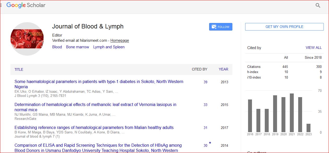
Journal of Blood & Lymph received 443 citations as per Google Scholar report