Research Article - (2024) Volume 10, Issue 1
Received: 30-Jan-2024, Manuscript No. jpnp-24-126366;
Editor assigned: 01-Feb-2024, Pre QC No. P-126366;
Reviewed: 14-Feb-2024, QC No. Q-126366;
Revised: 19-Feb-2024, Manuscript No. R-126366;
Published:
27-Feb-2024
, DOI: 10.37421/2472-0992.2024.10.282
Citation: Suruthelaya, T. M., Kaviya Suresh, K. K. Suriya
Prakaash and S. Nagalakshmi, et al. “An Effective Liposomal Gel Preparation for
the Long-lasting Herbal Mosquito Repellent.” J Pharmacogn Nat Prod 10 (2024):
282.
Copyright: © 2024 Suruthelaya TM, et al. This is an open-access article distributed under the terms of the Creative Commons Attribution License, which permits unrestricted use, distribution, and reproduction in any medium, provided the original author and source are credited.
Herbal mosquito repellent is a promising alternative to overcome the drawbacks of conventional mosquito repellents containing N, N-diethyl metatoluamide. The liposomal gel has a propensity for keratinizing the skin's horny layer and can penetrate deeper into the skin for enhanced absorption and is therapeutically effective and less toxic than other topical dosage forms, resulting in prolonged and controlled extract release. Vitex negundo (Chaste tree) leaves were dried and mechanically powdered. The powder was extracted with ethanol and distilled water (1:1), then the solvent was removed, dried using a lyophilization process, and stored. The phytochemical and composition analysis of the plant extract was done. The hydration process is carried out at a temperature of 40°C. Liposomal gel was prepared using the thin film hydration methods. A suitable base concentration was used to adjust the pH of the topical gel (6.5 –7.5). The liposomal dispersions were characterized for particle size distribution, SEM, XRD, ATR-FTIR, entrapment efficiency, in-vitro release study, and stability study. The liposomal gel was evaluated for colour, appearance, smoothness, Washability, skin irritation study, and Mosquito repellent activity. The developed formulations showed continuous extract release over 8 hours, extending the medication's residence time. The prepared optimized gel formulation was clear and stable after 90 days of storage under accelerated stability conditions. The mosquito-repellent activity of the optimized gel formulation was investigated using the arm-in-cage methodology. The field trials of the optimized gel were performed at 30, 60, 120, and 180 minutes, demonstrating excellent mosquito repellency. The total number of mosquitoes that landed was counted in a triplicate manner. The results of the present study revealed that Vitex negundo extract loaded mosquito repellent liposomal gel could produce fewer side effects, inexpensive, more effective, and long-lasting more than 10 hours to prevent mosquito-borne diseases like malaria, dengue, and chikungunya.
Vitex negundo leaves • Plant extract • Liposomal gel • Mosquito repellent activity • Long-lasting effect
Around the world, more than 700 million individuals are affected by various species of mosquitoes transmitting numerous diseases, and over one million deaths are reported annually across the globe. Mosquito-borne diseases are still a serious public health concern in humans [1]. Mosquitoes are responsible for transmitting more diseases than any other arthropod group. Various mosquito species belonging to the genera Anopheles, Culex and Aedes serve as vectors for diseases, including hemorrhagic fever, dengue fever, epidemic polyarthritis, yellow fever, chikungunya, malaria, Japanese encephalitis, filariasis, and other infections [2]. One of the most significant of these is malaria, which is caused by Plasmodium parasites and transmitted by female Anopheles mosquito bites, continues to produce skin irritations, allergic responses, and a significant illness impact in new-borns and young children in endemic areas. Despite taking several initiatives to prevent such transmission, many outbreaks continue. As a result, mosquito control and self protection from mosquito bites are now widely considered critical measures in the fight against mosquito-borne diseases [3]. Currently, the primary approaches for preventing mosquito-borne diseases are mosquito larvae control and human protection from mosquito bites through mosquito nets and repellents. Mosquito vaccines are an example of scientific advancements, although they are still in the beginning stages of development and are not yet recommended for human use [4]. In 1953, one of the most massively used synthetic repellents containing DEET (N,N–diethyl meta toluamide) was discovered to have a broad spectrum of activity against biting arthropods. To confront the issues, many people worldwide began purchasing DEET-based insect repellents. Everything was well until it was discovered that intake of the chemical component caused skin and mucous membrane irritation, poisoning with hyperammonemia and encephalopathy in children, urticaria syndrome, anaphylaxis, hypotension, and decreased heart rate. Mosquito coils are also well-known for being effective mosquito repellents. Pyrethrin, roughly 0.3-0.4 % of the coil mass, is the major active ingredient in mosquito coils. When we burn a mosquito coil, the insecticides evaporate with the smoke, preventing insects from entering the room. Many individuals, however, despise the scent of mosquito coils when burned because they believe they are dangerous to their health since they produce headaches, nausea, and dizziness. Mosquitorepellent liquidizers are also currently in widespread use throughout the world. Synthetic pyrethroids in mosquito-repellent liquidizers cause neurological damage if ingested accidentally. These have recently been detected as a source of hydrocarbon toxicity. The ample use of synthetic insecticides over the last five decades, on the other hand, has resulted in environmental risks and the development of physiological resistance in important vector species. Chemical-based mosquito repellents have excellent safety ratings, but they harm the human skin and nervous system, causing rashes, swelling, and eye irritations. These circumstances have prompted the development of natural mosquito repellents that are low-cost, effective, non-toxic, environmentally friendly, and biodegradable [5].
Liposomes are microscopic spheres with an aqueous core and one or more outer shells of lipids organized in a bilayer pattern. Liposomes offer various uses in medicines, cosmetics, and industries. Liposomes provide significant benefits over traditional dosage forms regarding drug loading, delivery, and sustained release. Liposomes are recognized as superior carriers because they can encapsulate and prevent hydrophilic and lipophilic drugs from degradation [6,7]. It also has a propensity for keratinizing the skin's horny layer and can penetrate deeper into the skin for enhanced absorption. Carriers that guarantee appropriate localization or penetration of the extract within or through the skin are used to formulate topical dosage forms to increase the local and limit the systemic effects or ensure proper percutaneous absorption. Liposomes are a solubilizing matrix for poorly soluble substances, a penetration enhancer, and a local depot when applied to the skin to minimize the adverse effects. Because liposomes seem to have a much higher diffusivity in the skin than most bare substances, they have been employed in transdermal drug delivery systems. As a result, topical liposome formulations may be more effective and less toxic than conventional formulations.
Although liposomes demonstrated promise for transdermal drug delivery, their practical application on the skin is minimal. However, these can be incorporated into gel that can be applied to the skin. [6] Liposomes incorporated into gel have been proven to be stable. Carbopol gel has been approved for pharmaceutical use in various administration routes, which might help maintain the optimum pH. Cutaneous use of these gel is ideal for application on the skin because they have good rheological characteristics, resulting in long residual periods at the site of administration, and they provide greater and prolonged skin concentrations than conventional gel [6,8].
Humans have been using the repellent activity of plant material for thousands of years, most easily by hanging crushed plants in houses, a technique that is still common in underdeveloped nations. Plants have also been used for ages as crude fumigants, in which plants were burned to drive away troublesome mosquitoes, and subsequently as oil formulations applied to the skin or clothing were first documented in ancient Greek, Roman, and Indian literature. Plant-based repellents are still widely used in this traditional manner throughout rural areas in tropical regions because they are the only defensive measure from mosquito bites available to many of the poorest communities, and some of these communities, like those in Europe and North America, prefer "natural" scented repellents because plants are considered as a safe and secure means of mosquito bite prevention [9,10]. Therefore, in search of herbal mosquito repellent to prepare a liposomal gel from Indian plants, we examined the leaves of Vitex negundo L., known to have the potential for pest control and mosquito repellency (Figure 1).

Figure 1. Pictorial representation of the developed formulation – Herbal mosquito repellent liposomal gel.
Collection, authentication and preparation of plant extract
The leaves of Vitex negundo L. were collected from the region of Siruvapuri, Thiruvallur district, Tamil Nadu, India [11,12]. The plant material was authenticated by P. Jayaraman, Director, Plant Anatomy Research Centre (PARC), Chennai, Tamil Nadu, India. A voucher specimen of the plant material was deposited in our department under PARC/2021/4534. Vitex negundo L. leaves were dried in the shade and processed into a coarse powder using a mechanical grinder. The powder was extracted with ethanol and distilled water (1:1). After extraction, the solvent was removed, dried using lyophilization, and stored in a container. Preliminary phytochemical analysis has been performed to identify the chemical constituents in the plant extract, like carbohydrates, glycosides, alkaloids, steroids, terpenoids, flavonoids, saponins, phenolic compounds, and tannins. The proximate composition analysis, water-soluble extractive, ethanol-soluble extractive, and ash concentration of plant extract have been done [12].
Analytical method development
10 mg (0.01 g) of plant extract was accurately weighed and transferred to a 10 ml volumetric flask, and the volume was adjusted to 10 ml with ethanol and distilled water (1:1) to give the concentration of 1000 μg/ml (stock solution A). From the standard stock solution (A), 1 ml was pipetted out in another 10 ml volumetric flask, and the volume was adjusted to 10 ml with ethanol and distilled water (1:1) to give the concentration of 100 μg/ml (stock solution B). Serial dilutions with concentrations 2, 4, 6, 8, and 10 μg/ml were prepared by transferring 0.2, 0.4, 0.6, 0.8, and 1.0 ml of the stock solution (B) in 10 ml volumetric flasks, respectively, and the volume was adjusted to 10 ml with ethanol and distilled water (1:1). The absorbance of resulting solutions was measured at 250 nm using UV-Visible Spectrophotometer (UV 1780, Shimadzu) and the standard calibration curve was constructed using Microsoft Excel 2007 program [12,13].
Preparation of liposomes
Thin film hydration is the most popular method for preparing multi lamellar liposomes. Soya lecithin and cholesterol were dissolved in 10 ml of methanol and chloroform (1:1), which was used as an organic solvent (solution of lipids). The plant extract was dissolved in 10 ml of ethanol and distilled water (1:1) and used as an extract solution. The procedure entails drying the organic solvent to the level where a thin film forms at the bottom of the rotary flash evaporator's round bottom flask. The organic solvent evaporates at 60°C for 30 minutes at 100 rpm. After evaporation, the thin film was dried overnight using a vacuumed oven. Then this thin lipid hydration layer suspension was dissolved in extract solution by vortexing for 10 minutes, and then it was allowed to hydrate at 40°C for 1 hr at 100 rpm. Then this liposomal suspension was sonicated for 3 minutes. The Liposomal final dispersions were stored in sterile glass vials at 4°C [12-14] (Tables 1 and 2).
| S. No | Ingredients | F1 | F2 | F3 | F4 | F5 | F6 |
|---|---|---|---|---|---|---|---|
| 1 | Soya lecithin | 35 mg | 25 mg | 22 mg | 20 mg | 18 mg | 16 mg |
| 2 | Cholesterol | 35 mg | 25 mg | 22 mg | 20 mg | 18 mg | 16 mg |
| 3 | Methanol | 5 ml | 5 ml | 5 ml | 5 ml | 5 ml | 5 ml |
| 4 | Chloroform | 5 ml | 5 ml | 5 ml | 5 ml | 5 ml | 5 ml |
| S. No | Ingredients | F1 | F2 | F3 | F4 | F5 | F6 |
|---|---|---|---|---|---|---|---|
| 1 | Plant extract | 10 mg | 10 mg | 10 mg | 10 mg | 10 mg | 10 mg |
| 2 | Ethanol | 5 ml | 5 ml | 5 ml | 5 ml | 5 ml | 5 ml |
| 3 | Distilled water | 5 ml | 5 ml | 5 ml | 5 ml | 5 ml | 5 ml |
Characterization of liposomal dispersions
Particle Size Distribution (PSD): Determination of particle size distribution of plant extract-loaded hydrated liposomal dispersions was carried out using a Dynamic Light Scattering (DLS) system (Malvern Zetasizer instrument). Liposomal dispersions were introduced to the sample dispersion unit containing a stirrer and stirred at 2000 rpm to minimize interparticle aggregation. For each formulation kept at 25°C, the mean particle size and polydispersity index were measured, and the polydispersity index will determine the size distribution of the liposome population. The size distribution of dehydrated liposomes (dry particles) was obtained with an optic microscope (Olympus BX60) connected to the computer with Image Pro-plus software [15-17].
Scanning Electron Microscopy (SEM): The surface and structure of the liposome were studied using Scanning Electron Microscopy (SEM). One drop of Liposomal dispersion was placed on the stab, which was then covered with clear glass (SEM sample holder) and dried in a desiccator. The sample was then sputter-coated with a gold coating of approximately 5 nm thickness and observed using a scanning electron microscope (FEG 200-High Resolution SEM). The photomicrographs were taken at a suitable magnification [17-19].
X-Ray Diffraction analysis (XRD): The crystallinity of the dried products was determined using X-ray Diffraction (XRD) analysis. Panalytical X’pert pro-X-Ray Diffract meter was used to record the powder XRD patterns of the samples at room temperature. To avoid particle orientation during preparation, samples were flattened on the sample holder. The following were the measuring conditions: Cu Kα radiation (λ=1.5406 Å), scanning angle 2θ ranged from 10°C to 90°C and scanning time was 3 minutes. During operation, the current and voltage were 30mA and 45kV, respectively [19,20].
Attenuated Total Reflectance-Fourier Transform Infrared Spectroscopy (ATR-FTIR): The functional groups of the samples were determined using ATR-FTIR spectroscopy. ATR-FTIR spectra of all the samples were measured on a Perkin Elmer FTIR spectrophotometer. Using an aqueous solvent group configuration that concentrates samples under vacuum conditions, the samples were thoroughly dried at 30°C. Once dried, the samples were examined by ATR-FTIR spectroscopy using a diamond crystal sample holder. Scanning was done with a range of 4000 to 600 cm-1 wavenumber and a resolution of 4 cm-1. OPUS software (version 4) was used to analyse the data, which was graphed as the reciprocal of wavelength (i.e., wavenumber or cm-1) [16,18,20].
Determination of Entrapment Efficiency (EE): Using the centrifugation method, plant extract associated with liposomes was isolated from the unentrapped extract. To determine the entrapment efficiency of liposomal dispersion formulations, 2 ml of the liposomal dispersions were centrifuged at 1000 rpm for 30 minutes at a controlled temperature of 4°C to isolate liposomes from the unentrapped extract. Using a UV-visible spectrophotometer (UV 1780, Shimadzu) against ethanol and distilled water (1:1), the supernatant containing unentrapped extract was measured at 250 nm. The following calculation was used to estimate the percentage of extract entrapment in liposomal dispersion formulations [17,20].
% Extract entrapment=(Total extract – Extract in supernatant liquid)/Total extract × 100
In-vitro release study of the liposomal dispersions
The In-vitro release of extract from the liposomal dispersions was studied using the Franz diffusion cell apparatus, consisting of a 3.4 cm inner diameter cylindrical glass tube that was opened at both ends (Donor chamber). As a diffusion medium, a freshly prepared homogenous mixture (ethanol and distilled water in a 1:1 ratio) was utilized. 2 ml liposomal dispersions were pipetted over the surface of the cellophane membrane (which had been soaked in the medium for 24 hours), and affixed to the tube's one end (bottom end). The entire assembly was positioned such that the bottom end of the tube containing liposomal dispersion just contacts (1-2 mm deep) the surface of the diffusion medium contained in another cylindrical glass tube that was opened at one end (Receiver chamber). The entire setup was placed on a magnetic stirrer and maintained at room temperature. The contents were stirred using a magnetic bar at 50 rpm for 8 hours, and 8 ml of samples were withdrawn at different time intervals and replaced with an equal volume of fresh diffusion medium. The samples were analyzed at 250 nm using a UV-visible spectrophotometer (UV 1780, Shimadzu) [12].
Stability study
Stability studies were conducted to assess for plant extract leaking from the liposome during storage. The optimized liposomal dispersion formulation (F2) was stored in a glass vial, closed with a grey butyl rubber closure, and sealed with aluminium foil at refrigeration temperature (4°C ± 2°C) and room temperature (25°C ± 2°C /60% ± 5% RH) for 90 days. Samples from the liposomal dispersion formulation (F2) stored for analysis were withdrawn at specific time intervals. Using a UV-visible spectrophotometer (UV 1780, Shimadzu), the withdrawn samples were evaluated for % entrapment of extract at 250 nm [17,20].
Preparation of liposomal gel
The suitable amount of Carbopol 940 was weighed and slowly added to 90 ml distilled water under continuous stirring by a magnetic stirrer (to prevent the formation of inevitable aggregates). After adding a solid substance, the gel swelled under average stirring for about 12 hours or until fully swollen and translucent. To obtain homogeneous gel dispersion, other ingredients, such as 15% w/v polyethylene glycol-400 (PEG-400), 0.5% w/v triethanolamine, methylparaben, and propylparaben, were added. Liposomal gel formulations were developed by mixing the 10 ml liposomal dispersion formulation with the gel. The final Liposomal gel formulations were stored in sterile bottles [12,21] (Table 3).
| S. No | Ingredients | G1 | G2 | G3 |
|---|---|---|---|---|
| 1 | Liposomal dispersion | 10 ml | 10 ml | 10 ml |
| 2 | Carbopol 940 | 0.5 g | 0.75 g | 1.0 g |
| 3 | Polyethylene glycol – 400 (PEG-400) | 2 ml | 2 ml | 2 ml |
| 4 | Triethanolamine | 3 drops | 3 drops | 3 drops |
| 5 | Methyl paraben | 0.1 g | 0.1 g | 0.1 g |
| 6 | Propylparaben | 0.05 g | 0.05 g | 0.05 g |
| 7 | Distilled water | 90 ml | 90 ml | 90 ml |
Evaluation of liposomal gels
Physical evaluation: The colour and transparency of the prepared liposomal gel formulations were assessed visually. The gel's smoothness and homogeneity were assessed by rubbing the formulations between the fingers [21,22].
Washability: The Washability of liposomal gel formulations was tested by first applying the gel to the skin and then assessing the ease and extent of washing it with distilled water while manually observing the effect [22].
Skin irritation: Human volunteers were used in this test. Three volunteers were chosen to apply the liposomal gel formulations, and the test was conducted after their informed assent was obtained. The 0.5 g of prepared liposomal gel formulations were applied to normal skin in a 6 cm2 area and then covered with a semi-occlusive bandage for 1 hour. The bandage was removed after the application period, the applied gel was scraped off thoroughly, and the region was visually examined for any rashes or other symptoms [22].
pH measurement: A digital pH meter (LMPH–12 model) was used to determine the pH of the prepared liposomal gel formulations. The electrode was dipped in the gel, and readings were recorded from the pH meter to confirm that the formulations did not cause any skin irritation to the patient after administration. For each formulation, the pH was measured thrice, and the average value was recorded [12,21].
Viscosity: The Digital Brookfield viscometer (DV2T model) was used to evaluate the viscosity of the liposomal gel formulations, using spindle number 64 at 10 rpm and a temperature of 25°C ± 2°C. 40 g of gel formulation was placed in a 50 ml beaker in such a way that it would allow the spindle groove to be dipped. The viscosity reading was measured after five minutes, and it was recorded. The same procedure was carried out three times, and the observations were averaged and recorded [22,23].
Extrudability: The process of filling the prepared liposomal gel formulations (20 g) into capped collapsible aluminium tubes and sealing them using a manual ointment sealing machine was used to determine their extrudability. The tubes (which contained different formulations) were placed in between two slides and securely clamped. After that, a 100 g weight was placed over the slides, and then the cap was opened, and the extruded ribbon length was measured after 10 minutes [22].
Spreadability: The spreadability of the prepared liposomal gel formulations was assessed by placing 1 g of each liposomal gel formulation on ground slides and sandwiching it with similar glide slides. To eliminate the entrapped air and establish a homogeneous film between the slides, a heavy mass was placed on the slides. From the edges, the excess gel content was scraped away. The following formula was used to calculate the spreadability of the gel formulations [22].
Spreadability (S)= (M × L)/T
Where M=Weight tide with upper slide; L=Length of the gel on the glass slide; T=Time taken (sec) to separate the glide slides from each other.
Swelling index: The swelling index of the developed liposomal gel formulations was evaluated by dissolving 2 g of each formulation in 10 ml of distilled water in separate beakers. The swollen formulations were withdrawn from the beakers and placed on various petri dishes after 1 hour. The weight of the contents was re-weighed, and the swelling index was calculated using the formula.
Swelling Index (SI)= [(Wt–Wo)/Wo] × 100
Where Wt=Weight of swollen gel formulation at time (t); Wo=Initial weight of gel formulation taken at time (zero) [22].
Extract content: To quantify the extracted content, 1 ml of each liposomal gel formulation was accurately measured and transferred to 100 ml of clean and dry volumetric flasks. Into the volumetric flasks, a homogeneous mixture (ethanol and distilled water in a 1:1 ratio) was added, the volume was adjusted to 100 ml with the same mixture, and it was sonicated for 5 minutes. Using a UV-visible spectrophotometer (UV 1780, Shimadzu), the absorbance of the solutions was measured at 250nm [23].
In-vitro release study of the liposomal gels
The In-vitro release of extract from the liposomal gel formulations was studied using the Franz diffusion cell apparatus, consisting of a 3.4 cm inner diameter cylindrical glass tube that was opened at both ends (Donor chamber). As a diffusion medium, a freshly prepared homogenous mixture (ethanol and distilled water in a 1:1 ratio) was utilized. 2 ml liposomal gel formulations were pipetted over the surface of the cellophane membrane (which had been soaked in the medium for 24 hours) and affixed to the tube's one end (bottom end). The entire assembly was positioned such that the bottom end of the tube containing liposomal gel formulation just contacts (1-2 mm deep) the surface of the diffusion medium contained in another cylindrical glass tube that was opened at one end (Receiver chamber). The entire setup was placed on a magnetic stirrer and maintained at room temperature. The contents were stirred using a magnetic bar at 50 rpm for an 8-hour, and 8 ml of samples were withdrawn at different intervals and replaced with an equal volume of fresh diffusion medium. The samples were analyzed at 250 nm using a UVvisible spectrophotometer (UV 1780, Shimadzu). To determine the order of release pattern and release rate constant, the acquired results were subjected to kinetic treatment [12,21].
Ex-vivo permeation study
A goat's ear pinna skin was collected and utilized in the permeation study. The skin was cleansed with cold water before, and a scalpel was used to remove full-thickness, non-dermatome skin, which was favoured as the membrane for the Franz diffusion cell apparatus. The skin permeation study was conducted using the Franz diffusion cell apparatus, which consists of a Donor chamber (a cylindrical glass tube with an inner diameter of 3.4 cm and an effective diffusion area of 0.785 cm2) and a Receiver chamber (12.0 ml capacity). The receiver chamber was filled with a homogeneous mixture (ethanol and distilled water in a 1:1 ratio). For a few minutes, the skin was immersed in the homogeneous mixture, and then it was placed between the donor and receiver chambers of the Franz diffusion cell, with the Stratum Corneum (SC) facing the donor chamber. In the donor chamber, 2 ml of the optimized liposomal gel formulation (G1) was pipetted. The entire setup was placed on a magnetic stirrer, and the receiver chamber was maintained at room temperature. The contents were stirred using a magnetic bar at 50 rpm for 8 hours, and 8 ml of samples were withdrawn from the receiver chamber at different intervals and replaced with an equal volume of a fresh homogeneous mixture prepared. The samples were analysed at 250 nm using a UV-visible spectrophotometer (UV 1780, Shimadzu) [24-26].
Apparent permeability coefficient
The freshly bought goat's ear pinna skin was utilized to calculate the apparent permeability coefficient using the formula,
Papp=(ΔQ/Δt) × 1/(A × CO × 60)
Where ΔQ/Δt=It is the flux over the ear pinna skin (μg/mL); A=It is the area of diffusion (cm2); CO=It is the initial extract concentration in the donor compartment (μg/cm3) and 60=it is a factor used to convert minutes into seconds. The amount of permeated liquid against time was plotted on a graph [23].
Accelerated stability study
According to the ICH guidelines, accelerated stability studies were conducted on the developed gel formulations. The ability of vesicles to retain the extract (i.e., extract retentive behaviour) was evaluated by storing the liposomal gel formulations at three different temperature conditions, i.e., 4°C ± 2°C (Refrigerator; RF), 25°C ± 2°C, 60% ± 5% RH (Room Temperature; RT) and 40°C ± 2°C, 75% ± 5% RH (Elevated Temperature; ET) for 90 days. The liposomal gel formulations were stored in glass vials, closed with grey butyl rubber closures, and sealed with aluminium foil. At certain intervals, samples of the optimized liposomal gel formulation were taken and analysed for the following essential parameters such as physical appearance, pH, viscosity, extrudability and extract content.
Mosquito repellent activity
Initially, volunteer’s forearms were properly cleansed with soap and dried completely. The left arm was used as a control, kept inside the mosquito cage containing 25 mosquitoes. In 20 seconds, the frequency of the vector landed on the forearm. The study began when the mosquitoes landed for more than 10 seconds. The arm was carefully removed from the mosquito cage after 20 seconds of time duration. The right arm, which served as the test, was smeared with the prepared optimized liposomal gel formulation (G1), and the trial was conducted at 30, 60, 120, and 180 minutes. The total number of mosquitoes that landed was counted in a triplicate manner. The percentage of mosquito repellence of the prepared optimized liposomal gel formulation was calculated using the formula below.
% Mosquito repellency= [(C – T)/C] × 100
Where C=Number of mosquitoes that landed on the control arm; T=number of mosquitoes that landed on the test arm [22,27].
The study of medicinal plant’s chemical constituents and active principles has gained much importance worldwide. Phytochemical analysis of Vitex negundo L. (plant extract) indicated the presence of glycosides, alkaloids, steroids, terpenoids, flavonoids, phenolic compounds, and tannins. The proximate composition analysis of Vitex negundo L. (plant extract), like watersoluble extractive, ethanol-soluble extractive, and ash concentration, has been presented in Table 5. The characteristic of the plant extract was found to have more solubility in water than ethanol (Tables 4 and 5).
| S. No | Chemical Constituents | Result |
|---|---|---|
| 1 | Carbohydrates (Benedict's test) | Absent |
| 2 | Glycosides (Treating with aqueous NaOH) | Present |
| 3 | Alkaloids (Mayer's test) | Present |
| 4 | Steroids (Salkowski test) | Present |
| 5 | Terpenoids (Chloroform and conc. H2SO4) | Present |
| 6 | Flavanoids (Lead acetate test) | Present |
| 7 | Saponins (Foam test) | Absent |
| 8 | Phenols (5% Ferric chloride) | Present |
| 9 | Tannins (Ferric chloride test) | Present |
| S. No | Parameters | Quantitative (%) |
|---|---|---|
| 1 | Total ash | 5.89 |
| 2 | Water soluble extractive | 18.74 |
| 3 | Ethanol soluble extractive | 11.67 |
The solubility of the plant extract (10 mg) in the homogeneous mixture (10 ml) was carried out, and it revealed that it is soluble in ethanol and distilled water in a 1:1 ratio. Based on the standard calibration curve of the plant extract in ethanol and distilled water (1:1), the linear regression equation (y=mx+c) obtained was y=0.01x+0.001
Where, y=Absorbance at the wavelength of 250 nm; m=Gradient of the line (slope); x=Concentration of the plant extract; c=its intercept with the y– axis. The regression coefficient was R2=0.996 (Figure 2).

Figure 2. Standard calibration curve.
Characterization of liposomal dispersions
A thin film hydration method with Soya lecithin and cholesterol was used to make liposomal dispersions containing plant extract, which were appropriate for developing liposomes without aggregation. The main objective of this work was to create multilamellar liposomes to improve extract permeability, improve therapeutic response, and minimize potential side effects.
Particle size distribution
The particle sizes of all liposomal dispersion formulations ranged between 629.7 nm and 686.6 nm. This method successfully produced a liposome population with a narrow size distribution. The results revealed that, with an increase in the concentration of soya lecithin and cholesterol, the particle size was found to be increased. The liposome population was heterogeneous, with a polydispersity index between 0.568 and 0.798. The polydispersity index values of the liposomal dispersion formulations show that the samples have a very broad particle size distribution. The optimized formulation (F2) has a mean particle size of 670.4 nm and a polydispersity index of 0.716, as shown in the size distribution by intensity graph (Figure 3 and Table 6).

Figure 3. Particle size distribution by intensity of the optimized liposomal dispersion formulation (F2).
| Formulation Code | Mean Particle Size (nm, ± SD) | Polydispersity Index (± SD) |
|---|---|---|
| F1 | 686.6 ± 14 | 0.798 ± 0.09 |
| F2 | 670.4 ± 12 | 0.716 ± 0.06 |
| F3 | 662.1 ± 18 | 0.621 ± 0.05 |
| F4 | 653.9 ± 10 | 0.607 ± 0.08 |
| F5 | 641.3 ± 15 | 0.579 ± 0.09 |
| F6 | 629.7 ± 11 | 0.568 ± 0.04 |
Scanning electron microscopy
SEM images show that the liposomes were formed in all the liposomal dispersion formulations. F1 and F2, which had higher proportions of soya lecithin and cholesterol, developed large, well-defined spherical vesicles, whereas F3, F4, F5, and F6 which had lower proportions of soya lecithin and cholesterol developed smaller, slightly deformed vesicles. These results reveal that soya lecithin and cholesterol have a significant role in liposome formation, shape, and size; formulations with a higher proportion of soya lecithin and cholesterol develop more spherical vesicles of larger size. The optimized liposomal dispersion formulation (F2) shows the existence of well-identified spherical vesicles of Vitex negundo L. plant extract in SEM photomicrographs (Figure 4).

Figure 4. Scanning electron microscopy images of optimized liposomal dispersion formulation (F2) - a) Measured at 100 μm and b) Measured at 3 μm.
X-Ray diffraction analysis
X-ray diffraction spectrum of the cholesterol and optimized liposomal dispersion formulation (F2) samples were done using a Panalytical X’pert pro-X-Ray diffractometer. The diffractograms were recorded under the following conditions: Voltage: 45kV, Current: 30mA, and room temperature. Those data were measured using scattering angle 2θ ranging from 10°C to 90°C. Furthermore, cholesterol and the prepared optimized formulation (F2) were compared with each other to verify whether it was in crystalline or in an amorphous state (Figure 5).

Figure 5. XRD spectrum of cholesterol and formulation F2.
Attenuated Total Reflectance-Fourier Transform Infrared Spectroscopy (ATR-FTIR)
This analysis identified the functional groups in the plant extract, soya lecithin, cholesterol, and the prepared optimized liposomal dispersion formulation (F2). Any interactions between the plant extract and lipid molecules were identified. Using a Perkin Elmer FTIR spectrophotometer, the ATR-FTIR spectrums of samples were measured from 4000 to 600 cm-1 wavenumber and had a resolution of 4 cm-1. Spectrum for plant extract, soya lecithin, cholesterol, and optimized liposomal dispersion formulation was compared (Figure 6 and Table 7).

Figure 6. ATR-FTIR spectra, a) Showed the spectrum of plant extract, b) Showed the spectrum of soya lecithin, c) Showed the spectrum of cholesterol and d) Showed the spectrum of optimized liposomal dispersion formulation (F2).
| Figure | Wave Number (cm-1) | Vibrations |
|---|---|---|
| Plant extract | 3282, 2924, 1617, 1369, 1266, 1029, 770 | O-H, C-H and C=C are stretching. C-H bending. C-O and C-C are stretching. C-H rocking |
| Soya lecithin | 2921, 2853, 1740, 1620, 1458, 1373, 1225, 1163, 1057, 861, 719 | C-H, C=O and C=C are stretching. C-H bending. C-O, C-N and C-C are stretching. C-H rocking |
| Cholesterol | 3393, 2921, 2850, 1736, 1644, 1461, 1371, 1232, 1129, 1052, 958, 884, 838, 799, 730 | O-H, C-H, C=O and C=C are stretching. C-H bending. C-O and C-C are stretching. C-H rocking |
| Optimized formulation F2 | 3286, 1641, 1456, 1087, 1015, 876 | O-H, C=O and C=C are stretching. C-H bending. C-O, C-N and C-C are stretching. C-H rocking |
Determination of Entrapment Efficiency (EE)
The entrapment efficiency of the liposomal dispersion formulations was determined by the centrifugation method. The entrapment efficiency of the liposomal dispersion formulations was between 35.07% and 49.16%. Formulation F2, with an EE of 49.16 % of plant extract, had the highest entrapment efficiency and formulation F6, with an EE of 35.07 % of plant extract, and had the lowest entrapment efficiency. According to the results, when soya lecithin and cholesterol were added to the mix, the % extract entrapment increased, but as the lipid concentration of both increased further, the % extract entrapment decreased (Figure 7 and Table 8).

Figure 7. Entrapment efficiency (%) of different liposomal dispersion formulations (F1, F2, F3, F4, F5 and F6).
| Formulation Code | Entrapment Efficiency (%, ± SD) | Cumulative Extract Release at the End of 8th hour (%, ± SD) |
|---|---|---|
| F1 | 46.08 ± 0.46 | 78.69 ± 0.76 |
| F2 | 49.16 ± 0.53 | 81.27 ± 0.39 |
| F3 | 42.89 ± 0.28 | 75.09 ± 0.45 |
| F4 | 39.93 ± 0.49 | 73.26 ± 0.57 |
| F5 | 36.27 ± 0.64 | 71.93 ± 0.23 |
| F6 | 35.07 ± 0.57 | 70.46 ± 0.61 |
In-vitro release study of the liposomal dispersions
The release studies of prepared liposomal dispersion formulations were carried out using the Franz Diffusion Cell apparatus. At room temperature, the experiment was carried out by removing 1 ml of the sample at each time interval over 8 hours and replacing it with an equivalent fresh diffusion medium. The sample was analysed spectrophotometrically at 250 nm; the % cumulative extract release was calculated for the prepared liposomal dispersion formulations F1-F6, as shown in Table 8. The % cumulative extract release was between 70.46 % and 81.27 % for all the developed formulations (F1-F6). Among all formulations, F2 seemed to have the highest % cumulative extract release of 81.27 %. The extract release patterns of all the formulations were shown to be increased over time (Figure 8).

Figure 8. % Cumulative extract release of the developed liposomal dispersion formulations (F1, F2, F3, F4, F5 and F6).
Stability study
Acceleration of extract leakage and increased vesicle size at higher temperatures indicated keeping the liposomal product refrigerated in storage stability analysis. Throughout the study, optimized liposomal dispersion (F2) was shown to be moderately stable in terms of aggregation, vesicle disruption, and fusion. The entrapment efficiency (%) for the extract content samples was calculated as a function of storage time. It may be concluded that the % entrapment of extract from the optimized formulation (F2) decreased slightly at room temperature. The results indicate that refrigerated liposomal dispersion reduces stability issues of liposomes (Figure 9).

Figure 9. Stability study of the optimized liposomal dispersion formulation (F2) at 4°C ± 2°C (refrigeration temperature) and 25°C ± 2°C, 60% ± 5% RH (room temperature) for a period of 90 days.
Evaluation of liposomal gel
Physical evaluation, washability and skin irritation: All three developed gel formulations (G1, G2 & G3) show a brown, translucent, homogenous, smooth-textured appearance with no solid particles found on rubbing between the fingers. The amount of Carbopol 940 in the gel formulations influenced the translucency considerably. A remarkable washability characteristic was observed when all the developed gel formulations were applied. Furthermore, no skin irritation, edema, rashes, or other dermatological symptoms occurred after 1 hour of gel formulation application (Table 9).
| Formulation Code | Color | Transparency | Smoothness | Washability | Skin Irritation |
|---|---|---|---|---|---|
| G1 | Brown | Translucent | Smooth | Good | No irritation |
| G2 | Brown | Translucent | Smooth | Good | No irritation |
| G3 | Brown | Translucent | Smooth | Good | No irritation |
pH measurement: The pH values of the developed gel formulations (G1, G2, and G3) were between 6.5 and 7.5, within the skin's normal pH range. These values are required for the gel to have a suitable viscosity and clarity. These pH values indicated that the gel formulations were unlikely to cause skin irritation. The conductivity values of gel formulations were constant. As a result, the prepared gel formulations were appropriate for topical application (Table 10).
| Formulation Code | pH | Viscosity (cp) | Extrudability | Spreadability (g.cm/sec) | Swelling Index (%) |
|---|---|---|---|---|---|
| G1 | 6.88 | 58,760 | +++ | 15 | 69.53 |
| G2 | 7.13 | 61,254 | ++ | 12 | 62.36 |
| G3 | 7.25 | 66,073 | ++ | 11.33 | 58.15 |
Viscosity: Because topical formulations are applied to the thin layers of the skin, homogeneity of the material is one of the most important characteristics, and gel viscosity plays an important role in regulating extract absorption. It's a crucial factor that affects pharmaceutical properties like spreadability and extrudability. The prepared gel formulations (G1, G2, and G3) have viscosity values ranging from 58.760 to 66.073 cps (Table 10). The results showed that when the concentration of polymer (Carbopol 940) increases, so does the viscosity of the gel formulations (Figure 10).

Figure 10. Viscosity of the developed gel formulations (G1, G2 and G3).
Extrudability: The extrudability of the developed gel formulations (G1, G2, and G3) was significant, with a considerable volume of extrudes ranging from ++ to +++ (Table 10). Extrudability decreases as the viscosity of the gel formulations increases, resulting in difficult extrusion from the collapsible aluminium tubes.
Spreadability: The prepared gel formulations must have good spreadability and satisfy the exemplar quality in topical application since the gel spreading aids in consistently applying the gel to the skin. The spreadability of gel determines their therapeutic effectiveness. The spreadability of the developed gel formulations (G1, G2 and G3) was 11.33-15 g.cm/sec (Table 10). The spreadability values indicated that a modest amount of shear might easily spread the gel formulations.
Swelling index: The swelling index indicated the matrix nature of the gel formulations, which facilitates a controlled extract release. The swelling index of the developed gel formulations (G1, G2, and G3) was determined to be between 58.15 % and 69.53 %, respectively (Table 10). G1 seemed to have the highest swelling index of 69.53 % of all the gel formulations.
Extract content: The extracted content in the prepared gel formulations (G1, G2, and G3) was measured at 250 nm using a UV-visible spectrophotometer. The gel formulations extract content was between 96.48 % and 97.59 % (Figure 11 and Table 11).

Figure 11. Extract content (%) of the prepared gels formulations (G1, G2 and G3).
| Formulation Code | Extract Content (%, ± SD) | Cumulative Extract Release at the End of the 8th hour (%, ± SD) |
|---|---|---|
| G1 | 97.59 ± 0.28 | 80.18 ± 0.36 |
| G2 | 96.81 ± 0.41 | 77.53 ± 0.59 |
| G3 | 96.48 ± 0.58 | 75.94 ± 0.61 |
In-vitro release study of the liposomal gel
The release studies of prepared gel formulations were carried out using the Franz Diffusion Cell apparatus. At room temperature, the experiment was carried out by removing 1ml of the sample at each time interval over 8 hours and replacing it with an equivalent fresh diffusion medium. The sample was analysed spectrophotometrically at 250 nm; the % cumulative extract release was calculated for the prepared gel formulations (G1, G2 and G3) as shown in (Table 11). The % cumulative extract release was between 75.94 % and 80.18 % for all the developed gel formulations. Among all gel formulations, G1 seemed to have the highest % cumulative extract release of 80.18 %. The extract release patterns of all the gel formulations were shown to be increased over time (Figure 12).

Figure 12. % cumulative extract release of the developed gel formulations (G1, G2 and G3).
In-vitro release kinetics [Theoretical calculation from % cumulative extract release]
The release data was fitted with various kinetic models to determine the extract release mechanism from the gel matrix. A kinetic analysis of the extract release data was performed by constructing a graph between % the cumulative extract releases and the square root of time. It suggested a linear correlation of extract release with time. Data obtained from in-vitro release studies was fitted with several kinetic models, including Zero order reaction, Higuchis Kinetics model, Korsmeyer Peppas reaction, First order reaction, and Hixson Crowell erosion equation methods, to analyse the release kinetics. The optimized gel formulation (G1) follows the Zero order reaction, with the regression coefficient (R2) value being the highest Figure 13 and Table 12. The values of regression coefficients confirmed the diffusion-dependent release of the extract (Figure 13 and Table 12).

Figure 13. In-vitro release kinetics –zero order reaction (Optimized gel formulation G1).
| Release Kinetics Models | Zero Order | Higuchi | Peppas | First Order | Hixson Crowell |
|---|---|---|---|---|---|
| R2 | 0.9955 | 0.0281 | 0.0190 | 0.9373 | 0.9667 |
Ex-vivo permeation study
The ex-vivo permeation study was done for the optimized gel formulation (G1) using the Franz Diffusion Cell apparatus. At room temperature, the experiment was carried out by removing 1 ml of the sample at each time interval over 8 hours and replacing it with an equivalent volume of a fresh homogeneous mixture. The study was carried out by graphing % cumulative extract release against the respective time shown in Figure 14. The gel formulation (G1) seemed to have the highest percentage extract release of 75.99 % at the end of 8 hours. The permeability coefficient of the optimized gel formulation (G1) was found to be Kp=0.0041cm/min (Figure 14).

Figure 14. Ex-vivo permeation study of % cumulative extract release for the optimized gel formulation (G1).
Accelerated stability study
According to ICH guidelines, the accelerated stability study was conducted on all the gel formulations (G1, G2, and G3). Gel formulations were stored in glass vials, closed with grey butyl rubber closures sealed with an aluminium foil at three different temperature conditions (i.e., 4°C ± 2°C (Refrigerator; RF), 25°C ± 2°C, 60% ± 5% RH (Room temperature; RT) and 40°C ± 2°C, 75% ± 5% RH (Elevated temperature; ET) for 90 days. The samples were taken at regular intervals and vortexed for 3-5 minutes in deionized water. According to the stability study, the prepared gel formulations were shown to be more stable at refrigeration temperature than at room temperature or elevated temperature. Around 90% of the extracted content was stable. Furthermore, the optimized gel formulation (G1) stored for stability studies underwent the following essential parameters: physical appearance, pH, viscosity, extrudability, and extract content, all exhibited good results even after 90 days, as shown in Table 14 (Tables 13 and 14).
| Formulation Code | G1 | G2 | G3 | |
|---|---|---|---|---|
| Extract content at 0th day (%, ± SD) | 25 °C | 97.59 ± 0.28 | 96.81 ± 0.41 | 96.48 ± 0.58 |
| Extract content at 30th day (%, ± SD) | 4 °C ± 2 °C | 97.41 ± 0.17 | 95.86 ± 0.31 | 95.39 ± 0.37 |
| 25 °C ± 2 °C, 60% ± 5% RH |
96.98 ± 0.21 | 94.89 ± 0.29 | 94.77 ± 0.47 | |
| 40 °C ± 2 °C, 75% ± 5% RH |
96.53 ± 0.24 | 94.54 ± 0.15 | 94.43 ± 0.14 | |
| Extract content at 60th day (%, ± SD) | 4 °C ± 2 °C | 97.35 ± 0.18 | 95.42 ± 0.28 | 95.17 ± 0.38 |
| 25 °C ± 2 °C, 60% ± 5% RH |
96.75 ± 0.14 | 94.45 ± 0.41 | 94.49 ± 0.29 | |
| 40 °C ± 2 °C, 75% ± 5% RH |
96.34 ± 0.28 | 94.28 ± 0.37 | 94.21 ± 0.41 | |
| Extract content at 90th day (%, ± SD) | 4 °C ± 2 °C | 97.13 ± 0.19 | 95.18 ± 0.27 | 95.01 ± 0.19 |
| 25 °C ± 2 °C, 60% ± 5% RH |
96.51 ± 0.27 | 94.23 ± 0.36 | 94.27 ± 0.35 | |
| 40 °C ± 2 °C, 75% ± 5% RH |
96.00 ± 0.25 | 94.08 ± 0.19 | 94.06 ± 0.28 | |
| Temperature | 4 °C ± 2 °C | 25 °C ± 2 °C, 60% ± 5% RH | 40 °C ± 2 °C, 75% ± 5% RH | |||||||||
|---|---|---|---|---|---|---|---|---|---|---|---|---|
| Regular Intervals | 0th day | 30th day | 60th day | 90th day | 0th day | 30th day | 60th day | 90th day | 0th day | 30th day | 60th day | 90th day |
| Physical appearance | Clear | Clear | Clear | Clear | Clear | Clear | Clear | Clear | Clear | Clear | Clear | Clear |
| pH | 6.88 | 6.87 | 6.85 | 6.84 | 6.88 | 6.86 | 6.84 | 6.81 | 6.85 | 6.83 | 6.81 | 6.78 |
| Viscosity | 58.760 | 58.746 | 58.714 | 58.692 | 58.760 | 58.717 | 58.685 | 58.667 | 58.760 | 58.703 | 58.659 | 58.641 |
| Extrudability | +++ | +++ | +++ | +++ | +++ | +++ | +++ | +++ | +++ | +++ | +++ | +++ |
| Extract content (%, ± SD) | 97.59 ± 0.28 | 97.41 ± 0.17 | 97.35 ± 0.18 | 97.13 ± 0.19 | 97.59 ± 0.28 | 96.98 ± 0.21 | 96.75 ± 0.14 | 96.51 ± 0.27 | 97.55 ± 0.28 | 96.53 ± 0.24 |
96.34 ± 0.28 | 96.0 ± 0.25 |
Mosquito repellent activity
The optimized gel formulation (G1) was examined using a mosquito cage for mosquito-repellent activity. The trial was conducted at 30, 60, 120, and 180 minutes in a triplicate manner. In the 0th minute, the optimized gel formulation (G1) had the maximum % mosquito repellence of 90.89 %, and it continued to function well until the 180th minute, when it reached 88.03 %. Various liposomal dispersion formulations (F1, F2, F3, F4, F5, and F6) were successfully developed utilizing the thin film hydration method to improve therapeutic response, increase extract molecule retention in the skin, and reduce the risk of side effects. Of all the developed liposomal dispersion formulations, F2 shows better entrapment efficiency and in-vitro extract release study results. Therefore, the optimized liposomal dispersion formulation (F2) was successfully incorporated into the Carbopol 940 to obtain gel formulations (G1, G2 and G3). Of all the developed gel formulations, the optimized gel (G1) presented good pH value, viscosity, Washability, extrudability, spreadability, swelling index, and no skin irritation. G1 also shows better results in extract content, in-vitro release study, and ex-vivo permeation study. Stability studies performed for optimized liposomal dispersion (F2) and gel (G1) indicate that the prepared liposomes have more stability at refrigeration than at room and elevated temperatures. Optimized gel formulation (G1) shows better results in mosquito repellent activity performed for about 3 hours (with regular time intervals) (Table 15).
| Formulation Code | % Mosquito Repellency (%, ± SD) | ||||
|---|---|---|---|---|---|
| 0th minute | 30th minute | 60th minute | 120th minute | 180th minute | |
| G1 | 90.89 ± 0.25 | 90.14 ± 0.21 | 89.84 ± 0.18 | 88.95 ± 0.29 | 88.03 ± 0.14 |
The prepared plant extract incorporated mosquito-repellent liposomal gel was successfully formulated using Carbopol 940. The liposomal delivery system is a one-of-a-kind technology that reduces adverse side effects and incompatibility caused by unacceptably high systemic absorption of extract and significantly increases the extract accumulation at the administration site. The liposomal delivery system offers better bioavailability, targeted cell delivery, and increased absorption. The above-performed characterizations and evaluations indicated that the developed gel formulation (G1) might prove to be a viable alternative to conventional mosquito repellents containing DEET (N, N-diethyl meta toluamide) in terms of fewer side effects, therapeutically more effective with added benefits of prolonged (long-lasting for more than 10 hours) and controlled extract release, which could lead to improved efficiency and better patient compliance. It is one of the most promising novel delivery systems in topical medications for successfully treating mosquito-borne diseases such as malaria, dengue fever, and chikungunya.
Conceptualization, S.T.M., K.S., N.S., G.S., S.J., and A.B.; Methodology, investigation and resources, S.T.M., K.S., S.P.K., N.S., N.A.C., G.S., K.P., and A.B.; Data curation, K.S., S.P.K., N.A.C., G.S., and A.B.; Writing-original draft preparation, S.T.M., K.S., S.P.K., A.B., K.P,; Writing-review and editing, N.S., N.A.C., G.S., and A.B.; Supervision, SN.S., N.A.C., G.S., S.J., and A.B.; Project administration, S.T.M., N.A.C., G.S., and A.B. All authors have read and agreed to the published version of the manuscript.
This research received no external funding.
Authors gratefully acknowledge the support from SAIF, IIT Madras, for PSD, SEM, XRD, and ATR-FTIR Characterizations. The authors are thankful to Sri Ramachandra Institute of Higher Education and Research (SRIHER), Porur, for providing the facility for the conduct of the research work. The authors would like to thank the Sri Ramachandra Institute of Higher Education and Research (SRIHER), Porur, for providing financial assistance.
The authors declare no conflict of interest.
Google Scholar, Crossref, Indexed at
Google Scholar, Crossref, Indexed at
Google Scholar, Crossref, Indexed at
Google Scholar, Crossref, Indexed at
Google Scholar, Crossref, Indexed at
Google Scholar, Crossref, Indexed at
Google Scholar, Crossref, Indexed at
Google Scholar, Crossref, Indexed at
Google Scholar, Crossref, Indexed at
Google Scholar, Crossref, Indexed at
Google Scholar, Crossref, Indexed at
Google Scholar, Crossref, Indexed at
Google Scholar, Crossref, Indexed at
Google Scholar, Crossref, Indexed at
Google Scholar, Crossref, Indexed at
Google Scholar, Crossref, Indexed at
Google Scholar, Crossref, Indexed at
Google Scholar, Crossref, Indexed at
Google Scholar, Crossref, Indexed at
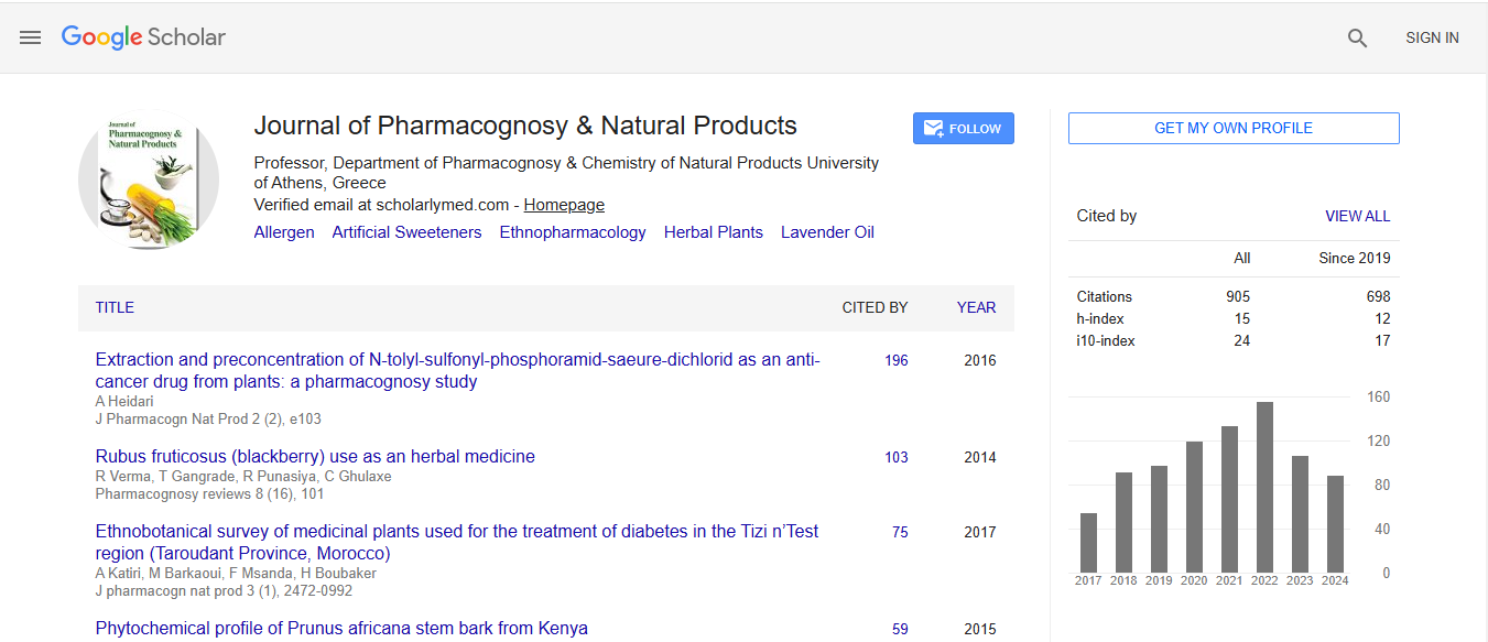
Journal of Pharmacognosy & Natural Products received 606 citations as per Google Scholar report