Mini Review - (2022) Volume 12, Issue 4
Received: 21-Nov-2022, Manuscript No. JBL-22-80569;
Editor assigned: 23-Nov-2022, Pre QC No. JBL-22-80569;
Reviewed: 07-Dec-2022, QC No. JBL-22-80569;
Revised: 14-Dec-2022, Manuscript No. JBL-22-80569;
Published:
21-Dec-2022
, DOI: 10.37421/ 2165-7831.2022.12.295
Citation: Mishra, Shruti “Recent Advances in the Screening of Dengue by Newer Parameters from Hematological Analyzers: A Mini-Review" J Blood Lymph 12 (2022) 295.
Copyright: © 2022 Mishra S. This is an open-access article distributed under the terms of the creative commons attribution license which permits unrestricted
use, distribution and reproduction in any medium, provided the original author and source are credited.
Dengue is one of the most commonly encountered infectious diseases in tropical countries. The features overlap with other causes of Acute Febrile Illness, and a rapid diagnosis is required in most centers. The usual modalities of diagnosis are serological based, where NS1 antigen, IgM, or IgG antibodies are assayed. Fast card-based kits are available but are less reliable than their (enzyme-linked immunosorbent assay) ELISA-based counterparts. There has been a recent automation boom in all healthcare facilities sectors. Automated hematological analyzers are an integral part of it, providing much additional information other than blood counts. Among these research parameters are HFLC (High Fluorescence Lymphocyte Count) and CPD (Cell Population Data) provided by the Sysmex XN series. These have taken the interest of pathologists, and some research articles are available that analyze these parameters' utility in the expeditious diagnosis of dengue. Studies show that HFLC is increased in AFI and correlates with dengue. Some studies have drawn Receiver Operating Curves (ROC) for determining cut-off values to differentiate dengue from other AFI. Jayaram et al. calculated a cut-off of 1.35% with 82.8% sensitivity and 87% specificity. Chabbra et al. computed a cut-off of 1.75% with 52% sensitivity and 90% specificity, a positive predictive value (PPV) of 72%, and a negative predictive value (NPV) of 80%. They also did a regression analysis on CPD and found that LY-X, LY-Z, LY-WX, LY-WZ, and MO-X were independent predictors of dengue fever. Ningombam et al. had different cut-off values for NS-1 antigen-positive only, IgM antibody-positive and dual-positive dengue patients, of 5.2%, 3.2%, and 2.6%, respectively. These studies show promising results and can help manage dengue patients, especially in resource-constrained settings in endemic zones, leading to better managing of dengue patients.
Dengue • Automated hematology analyzers • Acute Febrile Illness (AFI) • Sysmex-XN
Dengue is one of the most commonly encountered causes of Acute Febrile Illness in tropical countries. Dengue is caused by four different serotypes of viruses belonging to the Flaviviridae family. It is transmitted by female mosquitoes of mainly the Aedes aegypti genus. These mosquitoes have strips on their bodies and are often called "tiger" mosquitoes. They are fearless daytime biters and breed in stagnant water. The incidence of dengue increases exponentially during the rainy seasons. In flood-prone areas, the prevalence and disease burden is high.
Dengue fever usually has three phases-febrile, critical, and convlescent-phase. The characteristic symptoms are severe pain in bones,joints, muscles, and an eye is quite common. The critical phase can be challenging for some patients, especially those with a history of dengue infection. It can quickly turn fatal also in many cases. Sever form can lead to Dengue Hemorrhagic Fever (DHF) or Dengue Shock Syndrome (DSS). The exact pathophysiology still needs to be clarified. There is massive production of various cytokines, chemokines, and activation of the complement system and immune system. It leads to damage to the vascular endothelium and platelets. Due to endothelial damage, there is plasma leakage leading to DSS. Rapid depletion in platelet counts leads to superficial bleeding from the skin and mucosal surfaces resulting in DHF.
The presentation of dengue fever can overlap with other causes of AFI like malaria, chikungunya, leptospirosis, Rickettsial diseases, and in the current scenario, COVID-19 also. In all these cases, rapid, cheap, and accessible screening methods are required to manage these patients better. While serological assays for NS1 antigen, IgM, and IgG antibodies can be done as a strip test and on an ELISA-based platform, the latter has more sensitivity and specificity. The gold-standard method is molecular confirmation on PCR platform and virus isolation. However, it is expensive, laborintensive, and only readily available in some centers [1].
Recently there has been a boom in the automation of various routine laboratory tests, including automated hematological analyzers. It has cut down on turnaround time and manual labor and provides different research parameters that, if used wisely, can revolutionize the healthcare sector. Sysmex XN series analyzers are such high-end analyzers that give information regarding white blood cells. They provide a parameter called HFLC (High Fluorescence Lymphocyte Count), an "Atypical Lymphocyte" (AL) type where the cell cytoplasm has increased fluorescence. It is often postulated due to an increase in viral load in the cytoplasm of these lymphocytes. Numerous studies have been conducted on the RNA morphology of these lymphocytes compared to plasmacytoid lymphocytes. The high content due to the viral infection takes up the basic component of the Romanowsky stains and appears blue. These RNA content are also responsible for the increased fluorescence in these cells, similar to reticulocytes and immature platelets [2-5].
Other research parameters like CPD (Cell Population Data) are also available in the Sysmex XN series. These data denote the scatter position of the cells on the three positional axes, "X," "Y," and "Z." X-axis represents the internal complexity or granularity, also called Side Scatter (SSC), the Y-axis denotes the fluorescence intensity or the immaturity of the cells also known as Side Fluorescence (SFL). The Z-axis renders the size of the cell, also known as Forward Scatter (FSC). Whenever the RNA or DNA content in the cell cytoplasm increases, the SFL increases, like in the plasmacytoid lymphocytes/ reactive lymphocytes seen in dengue patients [5,6].
HFLC, even though increased in dengue, is not exclusive to this disease as reactive lymphocytes/ plasmacytoid lymphocytes can be seen in many other infective causes. Therefore, a few studies have tried to find a cut-off value of HFLC for predicting dengue in patients presenting with Acute Febrile Illness. A brief report on the various studies and their findings is given in Tables 1and 2.
| Sl. No. | Authors and Year | Total number of samples | HFLC% |
|---|---|---|---|
| 1. | Oehadian et al. (2015) | 93-Dengue Patients 11-Leptospirosis 6-Enteric fever 28-Healthy Controls |
The %ALa and %HFLCb had AUCc of 0.87 (95% CI 0.70-1.03) and 0.89 (95% CI 0.78-0.99), respectively, for distinguishing dengue from leptospirosis. |
| 2. | Tantanate et al. (2018) | Group 1-AFI (Infective causes) [n=155] Group 2-Autoimmune disorder [n=18] Group 3-Malignant conditions [n=18] Group 4-Healthy controls or patients with diseases other than previous groups [n=55] |
The correlation between HFLC and microscopic AL counts was 0.865 and 0.893 for bsolute and percentage counts, respectively. Patients with infections had the highest HFLC. Most of those patients (67.7%) had dengue infections. |
| 3. | Raharjo et al. (2019) | 47-DHFd 48-Healthy Controls |
DHF- 2.0-32. 3% Healthy Controls- 0-1.4% |
Abbreviations: AL- Atypical Lymphocytes; HFLC: High Fluorescence Lymphocyte Count; AUC: Area under the curve; DHF: Dengue Hemorrhagic Fever.
| Sl. No. | Authors and Year | Total number of samples | HFLC% (Cut-off) | Sensitivity | Specificity | AUC |
|---|---|---|---|---|---|---|
| 1. | Jayaram et al. (2021) | 445- Dengue cases 568- Controls (Dengue negative AFI) 449- Healthy Controls |
1.35 | 82.8 | 87 | - |
| 2. | Chhabra et al. (2021) | 97- Dengue Cases 202- Dengue Negative AFI 100- Normal controls |
1.75 (Dengue vs. other AFI) | 51.5 | 90.1 | 0.742 |
| 0.65 (Dengue vs. Healthy control) | 63.9 | 97 | 0.845 | |||
| 3. | Ningombam, et al. (2022) | 93- NS1Ag Positive 163- Dengue IgM Positive 36- Dual Positive 76- Dengue Seronegative |
>5.2 (NS-1 Ag Positive vs Negative controls) | 79.5 | 98.6 | 0.919 |
| >3.2 (IgM Positive vs. Negative controls) | 83.4 | 98.6 | 0.924 | |||
| >2.6 (Dual Positive vs. Negative controls) | 86.1 | 96 | 0.963 |
Reactive lymphocyte changes are mainly attributed to viral infections, autoimmune conditions, drug sensitivity, etc. The pathogenesis of dengue involves the activation of T-lymphocytes which leads to the downstream activation of B-lymphocytes and the production of various cytokines. The mediators cause an increase in the permeability of the blood vessels leading to the loss of fluid into the extra-capillary regions and activating the clotting cascade. These lymphocytes appear large with bluish cytoplasm on morphology and are flagged as "atypical lymphocytes" by the analyzers. As can be seen from Table 1, there is a strong correlation between HFLC and Atypical Lymphocytes.
Oehadian et al. were the first to study the use of HFLC and AL in tropical fevers in 2012 [3]. They used Sysmex- XE series for their analysis. They found no statistical difference in the total lymphocyte counts between dengue, leptospirosis, and enteric fever patients. However, the percentage of AL and HFLC was higher in patients with dengue than in those with leptospirosis and enteric fever. But none of these parameters correlated with the severity of the dengue infection. They drew Receiver Operating Curves (ROC) for AL% and HFLC%. The area under the curve (AUC) for AL was 0.87 (95% Confidence Interval [CI] 0.70-1.03) and for HFLC was 0.89 (95% CI 0.78-0.99), respectively, for distinguishing dengue from leptospirosis. Since they had only six enteric fever cases comparison couldn't be made with them [3].
Tantanate et al. studied 302 samples using Sysmex XN-3000 [7]. They divided the cases into four groups and found that the correlation between HFLC and microscopic AL counts was 0.865 and 0.893 for absolute and percentage counts, respectively. Patients with infections had the highest HFLC. Most of these were dengue patients (67.7%). Raharjo et al. used a Sysmex XN-1000 hematology analyzer to assess 48 healthy adults and 47 patients with DHF [8,9]. Dengue Hemorrhagic Fever (DHF) patients showed an increased HFLC% [2.0-32.3%] with a mean value of 11.5%. The average mean HFLC% was [0.0-1.4%] 0.3%.
Automated analyzers provide the results rapidly; these parameters are additional information retrieved without additional running costs. The cut-off values of HFLC% have been calculated in various studies, which helps differentiate dengue from other AFI. A cut-off of 1.35% was found by Jayaram et al., with a sensitivity of 82.8% and specificity of 87% [9]. They compared the HFLC values between 445 dengue-positive cases with 568 other patients of AFI on the Sysmex XN-9000 platform. Chabbra et al. calculated a cut-off of 1.75 with 52% sensitivity and 90% specificity, a Positive Predictive Value (PPV) of 72%, and a Negative Predictive Value (NPV) of 80% [10-12]. The number dengue positive cases included were 97, and dengue negative cases were 202. The analysis was done on Sysmex XN-1000 hematological analyzer.
Recently published data by Ningombam et al. had different cut-off values for NS-1 antigen-positive only, IgM antibody-positive and dualpositive dengue patients, of 5.2%, 3.2%, and 2.6%, respectively [11]. They analyzed 93 NS1Ag positive dengue patients, 163 patients with Dengue IgM positivity, 36 patients with dual positivity, i.e., both NS1 antigen and IgM antibody positivity, and 76 patients who were dengue seronegative. They processed their samples in the Sysmex XN-1000, which included the Sysmex XN-10 AHA module with a sampler.
One of the salient features of automated analyzers is the plots generated after the analysis of each sample. These plots are dynamic and give a representation of the individual cell features. The scatter plots are very useful in visually analyzing the composition of WBCs and, to a large extent, the morphological characteristics of these cells. The X-axis in Sysmex XN series denotes the Side Scatter (SSC), which depends on the cytoplasmic granularity and nuclear complexity like lobation. The more granular a cell and the more lobated the nucleus will make those cells lie farther away from the origin. Similarly, the Y-axis, which denotes the Side Fluorescence (SFL) of the cells, will have more immature cells or cells with increased fluorescence away from the origin. In the case of dengue, where the fluorescence of the cytoplasm increases due to viral load, cells will go up from the region where lymphocytes usually lie (Figure 1).

Figure 1.Scatter-plot showing upward trend (denoted by arrow) of the lymphocytes along Y-axis due to increased cytoplasmic fluorescence. Note: Color code in plot: Pink- Lymphocytes, Green- Monocytes, Light Blue- Neutrophils, and Red- Eosinophils.
These positional parameters have values used for research purposes only and are called Cell Population Data (CPD). Chabbra et al. had additional data on the CPD also. The most significant differences were observed in the lymphocytic and the monocytic CPD, which included LY-X, LY-Y, LY-Z, MO-X, MO-Y, and the MO-Z and the corresponding width of dispersion for lymphocytes (WX, WY, and WZ) with p-value <0.001. After regression analysis, it was found that LY-X, LY-Z, LY-WX, LY-WZ, and MO-X are independent predictors of dengue fever [10,11].
Further studies are required to correlate HFLC and CPD with other parameters like time of presentation and day of sample collection before its use as a routine screening parameter for dengue fever. But preliminary data from the above studies show that HFLC can be an easy, cost-effective, and time-saving tool for screening dengue patients, early identification, and management of these patients. These results are helpful, particularly in resource-constrained settings in endemic zones, primarily in developing nations, where the initial sample could assist screen patients for dengue fever and prompt the initiation of therapy with a better outcome.
[Crossref] [Google Scholar] [Pubmed]
[Google Scholar] [Pubmed]
[Crossref] [Google Scholar] [Pubmed]
[Google Scholar] [Pubmed]
[Crossref] [Google Scholar] [Pubmed]
[Crossref] [Google Scholar] [Pubmed]
[Crossref] [Google Scholar] [Pubmed]
[Crossref] [Google Scholar] [Pubmed]
[Crossref] [Google Scholar] [Pubmed]
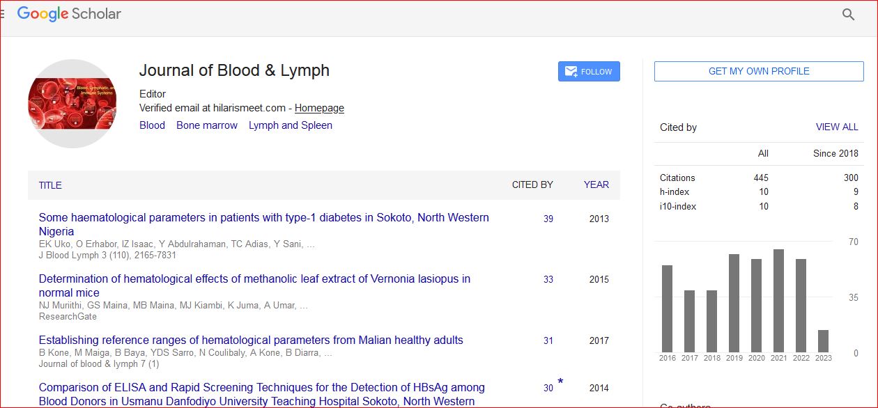
Journal of Blood & Lymph received 443 citations as per Google Scholar report