Mini Review - (2023) Volume 13, Issue 1
Received: 02-Jan-2023, Manuscript No. jbpbt-23-92635;
Editor assigned: 04-Jan-2023, Pre QC No. P-92635;
Reviewed: 16-Jan-2023, QC No. Q-92635;
Revised: 21-Jan-2023, Manuscript No. R-92635;
Published:
28-Jan-2023
, DOI: 10.37421/2155-9821.2023.13.557
Citation: Kim, Jaehi. “Living Things Abdominal Venous Extracellular Organelle in vitro Trackers by Applying Biotechnology Methods.” J Bioprocess Biotech 13(2023): 557.
Copyright: © 2023 Kim J. This is an open-access article distributed under the terms of the Creative Commons Attribution License, which permits unrestricted use, distribution, and reproduction in any medium, provided the original author and source are credited.
Despite the drawbacks of using conventional organic fluorescence probes, such as the weak fluorescent signal against the strong cellular autofluorescence background coupled with a fast-photobleaching rate, single-particle tracking (SPT) is a powerful method for exploring singlemolecule dynamics in living cells or tissues. Quantum dots (QDs), which enable tracking targets in multiple colours, have been suggested as an alternative to conventional organic fluorescence dyes. However, due to their hydrophobicity, cytotoxicity, and blinking issues, QDs are not ideal for applying SPT. The silica-coated QD-embedded silica nanoparticles (QD2) used in this study's improved SPT method have brighter fluorescence and are less toxic than single QDs. The label was kept after treatment with QD2 in 10 g/mL for 96 hours with impaired cell function, such as impaired angiogenesis, labelling efficiency was 83.76 percent. QD2's increased stability makes it easier to observe in situ endothelial vessel formation without needing to stain in real time. For 15 days at 4 °C, a cell maintain their QD2 fluorescence signal without experiencing significant photobleaching, proving that QD2 has overcome the limitations of SPT and is now capable of long-term intracellular tracking. With its superior brightness, photostability, and biocompatibility, QD2 was shown to be a viable alternative to single quantum dots or conventional organic fluorophores for SPT.
Fluorescence • Conventional • Cytotoxicity • Organic
Recently, the behaviour of targets in living cells or tissues was examined using single-particle tracking (SPT) analysis. For this type of imaging using total internal reflection fluorescence microscopy (TIRFM) or confocal laser scanning microscopy (CLSM), fluorescent probes are necessary. SPT probes have traditionally been made of organic fluorescence dyes like rhodamine b isothiocyanate (RBITC) or fluorescein isothiocyanate (FITC). These organic dyes do, however, have low brightness, high photobleaching, and hydrophobic properties. When stimulated by light, quantum dots (QDs), which are semiconductor nanocrystals, can emit light of a particular wavelength. By adjusting QDs' sizes and shapes, it is possible to create materials that emit light in a range of spectrums, from ultraviolet to infrared. The same method can be used to change the wavelength of light that stimulates QDs [1].
Additionally, harmful metals like cadmium, which makes up a sizeable portion of the most popular QDs, can interfere with bioimaging applications due to cytotoxicity. Although the distillation of even though single QDs can get around these issues, their blinking nature makes them unsuitable for real-time SPT. In a previous article, we described the creation of silica-coated QD-embedded silica nanoparticles (QD2), in which numerous QDs were incorporated into the silica template's surface and then enclosed in silica shells. QD2 has a number of benefits over single QD, including strong signal generation, low toxicity, and biocompatibility [2].
in order to get to the cellular organelles. By starving the cell for an hour and then giving it fresh growth or differentiation medium, the process can be sped up. Due to their noninvasiveness, which enables continuous vasculature monitoring, QD has been widely used to label and trace vasculatures in various tissues and is a potential tool to investigate the pharmacokinetics of drug candidates. In this regard, it is important to understand how endothelial cells react when exposed to QD directly. However, hydrophobic ligands are typically coated on QDs during fabrication for colloidal stability, which makes it simple for them to aggregate in physiological settings [3,4].
Because there are more QDs embedded in QD2, it emits a fluorescence signal that is 200 times stronger than a single QD without significantly blinking. Additionally, because of its hydrophilic property and ability to stop cadmium ions from leaking into the environment, the outer silica shell improves biocompatibility while reducing cytotoxicity. In addition, QDs of QD2 were shielded from the outside environment with In this study, QD2, a promising candidate for fluorescence probes of SPT with its excellent optical and biological properties, was tried for fluorescence imaging of human umbilical vein endothelial cells (HUVECs) via the SPT method [5].
In this study, QD2, a promising candidate for fluorescence probes of SPT with its excellent optical and biological properties, was tried for fluorescence imaging of human umbilical vein endothelial cells (HUVECs) via the SPT method. Prior to imaging HUVECs with fluorescence, QD2's optical characteristics and cytotoxicity towards them were examined. An ideal concentration for imaging was discovered. Fluorescence imaging was used to observe and compare the behaviours and cellular processes of QD2 uptaken HUVECs with those of native HUVECs following treatment of HUVECs with an optimised concentration of QD2. Long-term storage tests and a comparison with single QDs were also done to demonstrate the superiority of QD2 as a probe for SPT [6].
Prior to imaging HUVECs with fluorescence, QD2's optical characteristics and cytotoxicity towards them were examined. An ideal concentration for imaging was discovered. Fluorescence imaging was used to observe and compare the behaviours and cellular processes of QD2 uptaken HUVECs with those of native HUVECs following treatment of HUVECs with an optimised concentration of QD2. Long-term storage tests and a comparison with single QDs were also done to demonstrate the superiority of QD2 as a probe for SPT [7].
They are brighter, stable, and photostable than conventional organic dyes, making them an alternative probe for real-time cell tracking. Additionally, they offer a compelling alternative to current imaging technologies due to their cost-effectiveness and potential for future performance enhancements. Our research received CEFO IRB Council's approval in accordance with the Helsinki Declaration. A healthy donor's umbilical cord was used to isolate and culture human umbilical vein endothelial cells (HUVEC) in CEFOgroTMENDO. Before QD2 labelling, cells were starved for 1 h in basal media. The growth medium with QD2 was then substituted, and cells were incubated for 24 h at QD2 concentrations of 15 g/mL. Cells were cultured for live imaging, flow cytometry, and additional passaging after being washed.
None.
There is no conflict of interest by author.
Google Scholar, Crossref, Indexed at
Google Scholar, Crossref, Indexed at
Google Scholar, Crossref, Indexed at
Google Scholar, Crossref, Indexed at
Google Scholar, Crossref, Indexed at
Google Scholar, Crossref, Indexed at
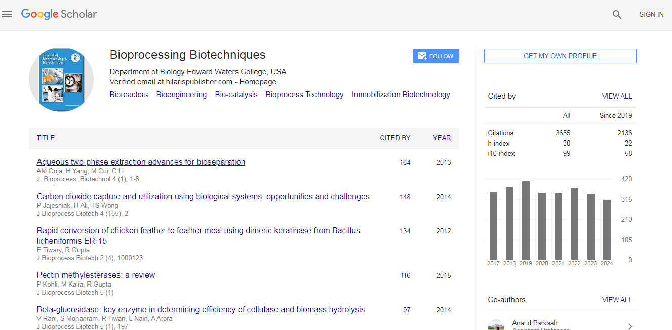
Journal of Bioprocessing & Biotechniques received 3351 citations as per Google Scholar report