Perspective - (2025) Volume 14, Issue 1
Received: 01-Feb-2025, Manuscript No. jme-25-169029;
Editor assigned: 03-Feb-2025, Pre QC No. P-169029;
Reviewed: 17-Feb-2025, QC No. Q-169029;
Revised: 22-Feb-2025, Manuscript No. R-169029;
Published:
28-Feb-2025
, DOI: 10.37421/2169-0022.2025.14.700
Citation: Maria, Balduin. “Influence of Surface Modification on Biocompatibility of Dental Implants.” J Material Sci Eng 14 (2025): 700.
Copyright: © 2025 Maria B. This is an open-access article distributed under the terms of the Creative Commons Attribution License, which permits unrestricted use, distribution and reproduction in any medium, provided the original author and source are credited.
Surface modifications can be broadly classified into physical, chemical and biochemical treatments. Physical modifications include methods such as grit blasting, sandblasting and plasma spraying, which create rough textures that enhance mechanical interlocking between the implant and bone tissue. These textures also influence cell behavior, promoting the attachment and proliferation of osteoblasts, the bone-forming cells. Roughened surfaces have been shown to increase Bone-To-Implant Contact (BIC) ratios, leading to better primary stability and faster healing times. Chemical treatments, such as acid etching and anodization, further modify surface energy and wettability, contributing to improved protein adsorption, which is crucial for early cell adhesion. Biochemical modifications involve immobilizing bioactive molecules, such as peptides, growth factors, or extracellular matrix proteins, on the implant surface to enhance specific cellular responses. These modifications aim to mimic the natural extracellular environment, guiding cell behavior at the molecular level. For example, coating the implant surface with Bone Morphogenetic Proteins (BMPs) or RGD peptides can stimulate osteogenic differentiation and accelerate osseointegration. These biofunctional surfaces also have the potential to modulate inflammatory responses and reduce fibrous encapsulation, which is often detrimental to implant stability. Advanced techniques like layer-by-layer assembly and self-assembled monolayers are increasingly being used to engineer such bioactive surfaces with precision and reproducibility.
Another critical aspect of biocompatibility is the implantâ??s resistance to bacterial adhesion, which plays a pivotal role in preventing peri-implantitis, a common cause of implant failure. Surface treatments such as silver ion implantation, titanium dioxide nanotubes, or antimicrobial peptide coatings aim to endow implants with antibacterial properties without compromising cytocompatibility. Nanostructured surfaces, in particular, have shown promise in promoting selective cellular responses supporting the growth of mammalian cells while inhibiting bacterial colonization. This dual-functionality approach addresses one of the most challenging aspects of dental implantology: achieving both high osseointegration and infection resistance.
Material characterization techniques like Scanning Electron Microscopy (SEM), Atomic Force Microscopy (AFM) and X-Ray Photoelectron Spectroscopy (XPS) are essential tools for analyzing the surface morphology, roughness and chemical composition of modified implants. In vitro studies involving human osteoblast-like cells (such as MG-63 or SaOS-2) are commonly used to assess cell viability, proliferation and differentiation on modified surfaces. In vivo animal studies further validate the histological and mechanical performance of implants under physiological conditions. Clinical data, although limited due to variability in patient factors and implant designs, generally support the conclusion that surface-modified implants demonstrate better early-stage integration and reduced healing times [2].
Google Scholar Cross Ref Indexed at
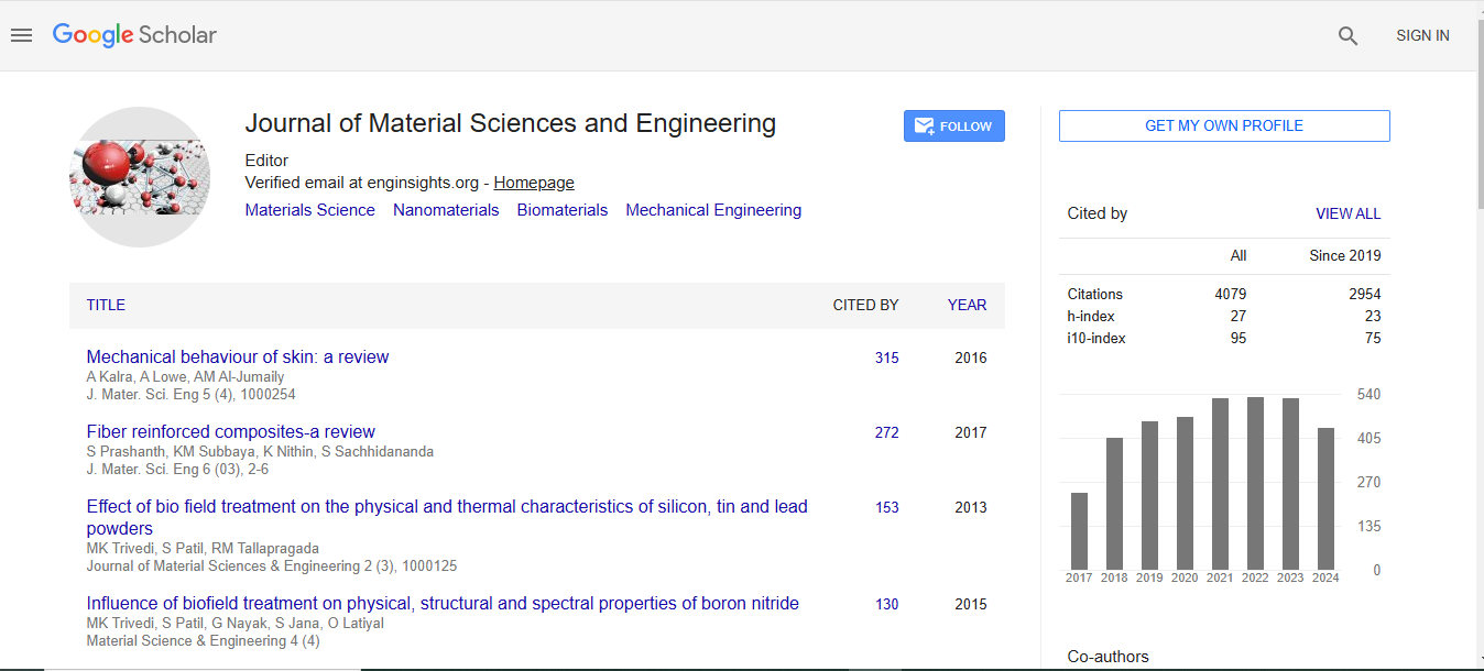
Journal of Material Sciences & Engineering received 3677 citations as per Google Scholar report