Research Article - (2022) Volume 0, Issue 0
Received: 07-Oct-2022, Manuscript No. JBL-22-76659;
Editor assigned: 13-Oct-2022, Pre QC No. JBL-22-76659;
Reviewed: 27-Oct-2022, QC No. JBL-22-76659;
Revised: 03-Nov-2022, Manuscript No. JBL-22-76659;
Published:
10-Nov-2022
, DOI: 10.37421/2165-7831.2022.12.016
Citation: Parhizgari, Najmeh, Farhad Rezaei, Mohammad-
Reza Khatami, Sayed Mahdi Marashi, et al. "Cytomegalovirus Viremia and
Risk Factors in Renal Transplant Recipients: A Case-control Study". J Blood
Lymph 12(2022): 016.
Copyright: © 2022 Parhizgari N, et al. This is an open-access article distributed under the terms of the creative commons attribution license which permits unrestricted use, distribution and reproduction in any medium, provided the original author and source are credited.
Background: In spite of antiviral prophylaxis regimens, Cytomegalovirus (CMV) remains a major reason for morbidity and allograft failure in kidney transplant recipients. This study aimed to investigate the incidence of early or late onset of CMV viremia in kidney transplant recipients and evaluate the correlation of laboratory findings and graft origin with CMV viremia.
Methods: In this prospective case-control study, 192 kidney recipients were evaluated for the timing and potential risk factors based on detectable CMV viremia (≥200 copies/ml) and all-correlates were assessed using multivariate logistic regression models.
Results: 153 participants from examined patients were eligible to enter the study. The risk of CMV viremia with viral loads ≥200 copies/ml was receiving a graft from a deceased donor. Importantly, CMV viremia mostly occurred 4 months after transplantation, while the patients were expected to be on CMV prophylaxis.
Conclusions: Receiving a renal graft from a deceased donor significantly raises the incidence of viremia in renal transplant patients. The median month of CMV viremia occurrence was month 4th after transplantation. Serum testing showed a significant increase in creatinine and a decrease in platelets in the CMV positive group compared to the control group. Our results indicated that the viremia has not affected the survival of the allograft or patient.
Renal transplant infection • CMV viremia • CMV Risk factors • CMV infection • Deceased donors
CMV: Cytomegalovirus; BKV: BK Polyomavirus; HHV6: Human Herpesvirus-6; EBV: Epstein Bar Virus; PCR: Polymerase Chain Reaction; RT: Renal Transplant; EDTA: Ethylenediaminetetraacetic Acid; SGOT: Serum Glutamic Oxalacetic Transaminase; SGPT: Serum Glutamic Pyruvic Transaminase; ALP: Alkaline Phosphatase; WBC: White Blood Cell; FBS: Fasting Blood Sugar; DNA: Deoxyribonucleic Acid; RNA: Ribonucleic Acid; IQR: Interquartile Range.
In spite of effective anti-viral drugs and risk-balanced prophylaxis regimen, Cytomegalovirus (CMV) infection is still considered as a major concern regarding to the morbidity of kidney transplant patients [1]. The prevalence of CMV infection in Iran is estimated to be 99% among people 18-64 years [2]. The risks of CMV-related complications in kidney transplant recipients vary depend on the type of transplant and whether the recipient and donors already had latent infections [3]. In Renal Transplant (RT) patients, infection occurs either primarily or as reactivation of latent virus depends on kidney donor or recipient CMV serostatus, however, most of CMV- reactivation in Iran categorized in the middle risk group [4]. Reactivation of CMV in kidney transplant recipients may manifest across a clinical spectrum from asymptomatic viraemia to tissue-invasive disease [5]. The reactivation can lead to CMV disease, the direct effects of the disease due to the attendance of high rates of viral replication and lytic virus production, or indirect effects including the virus interaction with the immune response, acute rejection, graft dysfunction, opportunistic infections, diabetes mellitus, and malignancies [6]. The indirect effects of CMV are assumed to be mediated by the production of cytokine and chemokine [7,8].
Re-emergence of CMV viremia could occur as an ‘early onset’ or ‘late onset’ infection [9]. Approximately, 10%-50% of allograft recipients get the early-onset CMV infection after or during receiving anti-viral prophylaxis. The clinical manifestations are developed in nearly half of these patients, and the recurrence is observed in up to 30% of the successfully treated cases. CMV infection appears within the first six months after organ transplantation, and recurrences happen during three months of the fulfillment of therapy of the initial episode. Late CMV disease is developed more than 6 months after organ transplantation, and it is often associated with the requirement to raise the immunosuppression level due to late episodes of rejection [10,11].
In some RT patients, the allograft origin may change the risk of CMV infection after transplantation. Several studies show that outcome of living donor kidney transplantation has been better than that of deceased donor kidney transplantation and the deceased donors might extend the infection risk through donor-derived nosocomial organisms or severe immunosuppression [12,13]. In a Cohort study in Europe, deceased donor transplantation was observed to be associated with increased incidence of CMV viremia [14].
According to our knowledge, there is no comprehensive study on correlation of laboratory findings, and graft origin with CMV viremia as well as late or early onset of CMV infection in kidney transplant recipients in Iran. We, therefore, conducted a prospective case-control analysis to examine the above-mentioned factors which could be affected by the virus viremia or cause an increase the risk of developing the infection among Iranian renal transplant patients.
Study design and setting
This study was a prospective case-control in which the recruited subjects comprised 153 RT patients had referred to Imam Khomeini Hospital (IKH), Tehran, Iran between February 2019 and February 2021. Figure 1 shows the overall flowchart of the prospective case-control study design. To select the case group, the suspected or randomly found patients with CMV infection were sampled and the control group was collected from RT patients with no typical symptoms of CMV infection from 192 patients who were examined and tested, 43 and 110 patients were recruited in the case and control group, respectively. The inclusion criteria included the adult patients (age ≥ 18 years) including outpatients and hospitalized RT patients who received a live or deceased donor kidney transplant in Imam Khomeini Hospital. All RT patients in both groups were donor-positive and recipient-positive, either with clinical symptoms (fever, low glomerular filtration rate, and signs of urinary tract infection) or asymptomatic. A viral load ≥ 200 copy/ml was defined as viremia to enter to the case group. PCR for determining BKV, HHV6, and EBV was performed on all the plasma samples of two groups and the negative results provided the qualification to enter the case or control categories (Figure 1).
The study was evaluated and approved by the Ethical Committee of Tehran University of Medical Sciences. Informed consent was obtained from all patients before specimen collection. The subjects’ general data (age, gender and months after transplantation, donor source, and date of transplantation) and underlying diseases which caused kidney loss of function were recorded in a questionnaire. First, 3 ml aliquot of whole blood sample was withdrawn from each participant, then plasma was separated immediately from 1 ml using EDTA as anti-coagulant and it was stored at -70˚C in RNase DNase free micro-tubes for viral nucleic acid extraction. Two ml of blood was sent to the diagnostic laboratory to determine laboratory parameters including white blood cell count, platelet count, serum FBS, serum creatinine, serum uric acid, Serum Glutamic Oxalacetic Transaminase (SGOT), Serum Glutamic Pyruvic Transaminase (SGPT), and Alkaline Phosphatase (ALP).
Immunosuppressive therapy
In IKH, most patients received routine triple immunosuppression with a calcineurin inhibitor (CyA/tacrolimus), mycophenolate sodium or mycophenolate mofetil, and oral prednisolone. The at-risk patients received thymoglobulin induction. Target tacrolimus trough levels were typically between 5 and 10 μg/L for the first 3 months, with the dose of prednisolone reduced to 5-10 mg daily by 3 months. Biopsy-proven T cell-mediated rejection episodes were managed by intravenous methylprednisolone, and T cell depleting antibody being prescribed for steroid-resistant rejection episodes. CMV prophylaxis with valganciclovir was administered for 100 days to patients.
DNA extraction and PCR for CMV, BKV, HHV6, and EBV
Viral nucleic acids were extracted from 200 ul of plasma samples using Roche viral nucleic acid extraction kit (Roche, Basel, and Switzerland), according to the manufacturer’s instruction. BKV, HHV6, and EBV infections were examined by conventional PCR as described before [15,16].
Statistical analysis
Descriptive statistics including means, standard deviations of quantitative variables, and frequencies (%) of qualitative variables were computed. Continuous and categorical variables were presented as median (IQR) and n (%), respectively. The Wilcoxon ranking tests, X2 test or Fisher's exact test were used to compare the differences among different groups, depending on the situation. The multivariate logistic regression models were used to control possible cofounders after analyzing based on the CMV PCR test. A two-sided α of less than 0.05 were regarded statistically significant. Statistical analyses were done using R version 4.0.3 (2020-10-10).
The study population included 100 patients and 100 controls. There was no significant difference between patients and controls with respect to the mean age, gender, and being a healthcare worker (P>0.05; Table 1). Univariate analysis revealed that Hepatitis B Virus (HBV) seropositivity was significantly associated with sharing shaving machines, sharing toothbrushes, unhygienic dental care, and living abroad (P<0.05). However, this univariate analysis did not show any significant differences between patients and controls regarding exposures to contaminated medical devices, endoscopy, blood transfusion, travel to endemic areas, major trauma unprotected sex, tattooing, and eating from common plates (P>0.05).
Based on the qPCR test and CMV viral load, 43 patients among all the 192 surveyed ones had more than 200 copies of the CMV genome in plasma. The study population included 43 patients and 110 controls. The univariate analysis revealed that those who had CMV positive test, showed significantly higher blood creatinine and lower palette. CMV infection was occurred significantly (P<0.05) more in patients who received the kidney from a deceased donor. However, this univariate analysis did not show any significant differences between patients and controls groups regarding the months after transplantation (Table 1). The main reason for the patient’s loss of function was blood pressure (72%) following diabetes (36 %) (Figure 2).

Figure 2. The frequency of kidney loss of function reasons in the investigated patients in both case-control patients.
Demographic profile of transplant recipients
The descriptive variables categorized by CMV molecular test results are presented in Table 1. Among the case-control participants, the mean age was 51.3 years. The majority of patients were males (71%), and primary renal disease was chronic glomerulonephritis in 57% of CMV- positive patients. About 27% of the kidney transplant recipients received the organ from living donors (Table 1).
| Characteristic | N1 | Overall (N=153) |
Negative (N=110) |
Positive (N=43) |
p-value2 |
|---|---|---|---|---|---|
| Sex | |||||
| Female | 153 | 44 (29%) | 31 (28%) | 13 (30%) | 0.8 |
| Male | 109 (71%) | 79 (72%) | 30 (70%) | ||
| Age | 153 | 54 (39, 61) | 55 (41, 62) | 45 (39, 60) | 0.2 |
| WBC | 151 | 7.60 (5.70, 9.30) | 8.00 (5.90, 9.50) | 6.70 (5.43, 8.75) | 0.1 |
| Platelets | 151 | 197 (164, 253) | 202 (172, 264) | 182 (153, 221) | 0.019 |
| FBS | 145 | 105 (89, 130) | 108 (89, 134) | 104 (84, 120) | 0.2 |
| Creatinine | 153 | 1.41 (1.12, 1.79) | 1.33 (1.11, 1.73) | 1.57 (1.31, 2.14) | 0.008 |
| Uric. Acid | 129 | 5.80 (5.00, 6.70) | 5.70 (4.93, 6.70) | 6.00 (5.60, 6.80) | 0.2 |
| SGOT | 90 | 20 (15, 25) | 21 (16, 25) | 20 (14, 26) | 0.4 |
| SGPT | 90 | 32 (19, 50) | 34 (19, 50) | 30 (20, 43) | >0.9 |
| ALP | 80 | 232 (178, 330) | 243 (183, 352) | 229 (178, 308) | 0.7 |
| *Months after transplantation | 153 | 5 (2, 15) | 5 (2, 22) | 4 (3, 7) | 0.4 |
| Survival | |||||
| Rejected | 153 | 2 (1.3%) | 2 (1.8%) | 0 (0%) | >0.9 |
| Survived | 151 (99%) | 108 (98%) | 43 (100%) | ||
| Donor sources | |||||
| Deceased donor | 153 | 112 (73%) | 73 (66%) | 39 (91%) | 0.002 |
| Living donor | 41 (27%) | 37 (34%) | 4 (9.3%) | ||
Note: 1N (%); Median (IQR);2 Pearson's Chi-squared test; Wilcoxon rank sum test; Fisher's exact test;*months after transplantation.
Risk Factors of CMV viremia
According to the multivariate logistic regression models, age, WBC, platelets, FBS, creatinine, uric acid, and donor source had p-values less than 0.2 and entered to multivariable analysis. The result of model indicates that receiving organ from a deceased donor significantly augments the risk of CMV infection in the case group (Table 2).
| Characteristic | OR | 95% CI | p-value |
|---|---|---|---|
| Age | 0.98 | 0.95, 1.02 | 0.3 |
| WBC | 0.92 | 0.75, 1.13 | 0.5 |
| Platelets | 0.99 | 0.98, 1.00 | 0.006 |
| FBS | 1 | 0.98, 1.01 | 0.4 |
| Creatinine | 0.69 | 0.25, 1.87 | 0.5 |
| Uric Acid | 1.06 | 0.76, 1.48 | 0.7 |
| Donor sources | |||
| Deceased donor | - | - | - |
| Living donor | 0.29 | 0.07, 0.96 | 0.057 |
Abbreviations: OR: Odds Ratio; CI: Confidence Interval.
While CMV serostatus has an important role in the CMV infection development in kidney transplant recipients, it is yet not clear whether other risk factors could have the same function. The present study aimed to investigate the risk factors which increase the CMV viremia occurrence in this group. Thus, the results of serum laboratory tests in CMV positive patients were compared to the results obtained from negative patients to find the significant different tests between two groups. Moreover, we investigate the incidence of early or late CMV infection, risk factors, and consequences of CMV viremia among kidney transplant recipients compared to the control patients.
The present results indicate that RT patients who received the allograft from deceased donors faced a higher risk of CMV viremia after transplantation mostly in the first four months. Recent analysis in RT patients suggests grafting from a cadaveric would raise the risk of CMV viremia.
CMV viremia in our study emerged generally 4 months after transplantation. In accordance with a cohort study in Europe, viremia emerged within the first three months after transplantation among 19.2% of CMV+ patients (14). Given acute rejection episodes occurred within the first month post-transplantation, the association with CMV infection in this period can be attributed to the excessive immunosuppression associated with acute rejection treatment. Infection recurrence probably promotes by the initial immunosuppression and cessation of anti-viral prophylaxis. It is suggested that, patients with a higher risk of CMV infection be treated by antiviral prophylaxis during this time. Our results showed that early CMV infection emerges in 4 months after transplantation although there was no significant difference in the time of CMV detection between both CMV+/- groups which may be considered as an alarming time to examine the presence of CMV viremia in the RT patients. The late-onset CMV infection has not been detected in late months. It was recorded on the month 7th of receiving the kidney among the investigated patients. In other studies, the development of late-onset CMV disease was reported in approximately 18% of patients even in the presence of either prophylactic strategy [17,18].
A delayed onset of infection may occur after the discontinuation of prophylaxis, and there is evidence of a lower CMV incidence after prophylaxis suspension following a long period of drug use [19]. According to the arguments in favor of prophylaxis, the use of antivirals would reduce the indirect effects of viral replication by preventing both viral replication and disease [20].
Unlike the proved fact in other studies, in the present study, CMV viremia did not significantly affect the allograft and patient’s survival [21]. Given we investigated seropositive RT patients and in patients who were CMV seropositive, viral replication occurs in the context of preexisting immunity, hence the observed replication rate is slower in such individuals. As a result of the widespread use of antiviral prophylaxis and preemptive therapy, the incidence and severity of CMV disease and its indirect effects are significantly reduced. The incidence of CMV in the renal transplant population is estimated to be between 8% and 32% [10]. After transplantation, patients in the CMV+ group displayed worse serum creatinine levels without significant differences in graft and patient survivals [8]. Raised Serum Creatinine and diminished platelets, are results which confirm the facts that CMV infection must be noted in all renal transplant recipient whose creatinine has been elevated, even asymptomatic recipients [22].
Our results did not investigate the local detection of CMV in the allograft, so it is important to consider that the mere detection of CMV does not essentially exclude the presence of CMV in the blood. Indeed, lack of serum CMV positive test does not completely rule out CMV infection in these patients, since transient periods of CMV viremia had been found in some cases due to the compartmentalized or localized CMV diseases [23,24]. In the present study, all subjects had received CMV antivirals including ganciclovir (1.25 mg/kg IV daily as induction for 1 month, which then was switched to oral valgancyclovir) or valcyte (450 mg, according to their plasma creatinine levels) for the first 3 months’ after transplantation. In the cases of CMV DNAemia, some patients did not show any typical syndromes of CMV infection [25,26].
In this study, we determined CMV viremia by detection of virus DNA in the patients’ blood. However, new diagnostic method based on the amplification of CMV RNA in blood samples has been commercialized, and we recommend that future studies categories the case-control patients based on these novel technique. Significant increase the incidence of CMV viremia in RT patients as a result of receiving a kidney from a deceased donor is known as a novel insight in Iran. No late CMV infection was detected in our case group and the viremia did not affect the survival of the allograft or patient. Future case-control studies are recommended to perform based on CMV diagnosis by viral late gene expression.
Ethics approval and consent to participate
The study was evaluated and approved by the Ethical Committee of Tehran University of Medical Sciences. Before specimen collection, informed consent was obtained from all patients.
Availability of data and materials
The datasets used and analyzed during the current study are available from the corresponding author on reasonable request.
Conflict of interest
The authors have no competing interest to declare. Funding sources had no role in the design and conduct of the study; collection, management, analysis, and interpretation of the data; and preparation, review, or approval of the manuscript.
Funding
This study was part of a PhD thesis in Medical virology supported by Tehran University of Medical Sciences (grant no. 46944-99-1-99).
Acknowledgments
We would like to thank all the patients who kindly participate in our study.
Author contribution
Conceived and designed the experiments: NP, FR, TMA. Performed experiments: NP, FB. Contributed materials/analysis: NP, MRK, SMM, MD, MZG, FAN. Analyzed the data: NP, MF. Wrote the paper: NP, FR, TMA.
[Crossref] [Google Scholar] [Pubmed]
[Crossref] [Google Scholar] [Pubmed]
[Crossref] [Google Scholar] [Pubmed]
[Crossref] [Google Scholar] [Pubmed]
[Crossref] [Google Scholar] [Pubmed]
[Crossref] [Google Scholar] [Pubmed]
[Crossref] [Google Scholar] [Pubmed]
[Crossref] [Google Scholar] [Pubmed]
[Crossref] [Google Scholar] [Pubmed]
[Crossref] [Google Scholar] [Pubmed]
[Crossref] [Google Scholar] [Pubmed]
[Crossref] [Google Scholar] [Pubmed]
[Crossref] [Google Scholar] [Pubmed]
[Crossref] [Google Scholar] [Pubmed]
[Crossref] [Google Scholar] [Pubmed]
[Crossref] [Google Scholar] [Pubmed]
[Crossref] [Google Scholar] [Pubmed]
[Crossref] [Google Scholar] [Pubmed]
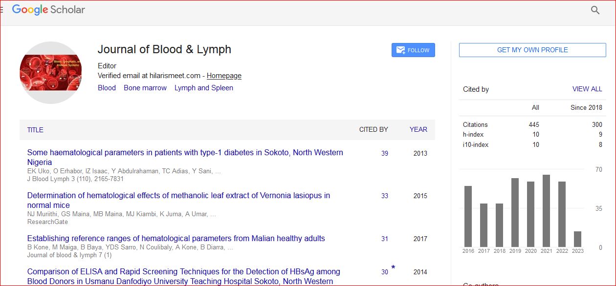
Journal of Blood & Lymph received 443 citations as per Google Scholar report