Short Communication - (2021) Volume 11, Issue 5
Received: 09-Sep-2021
Published:
20-Sep-2021
, DOI: 10.37421/2161-0991.21.11.190
Citation: Edward Thorp. “A New Technique of Heart Lesion in
Rat.” J Transplant Technol Res 11 (2021): 190.
Copyright: © 2021 Edward Thor. This is an open-access article distributed under
the terms of the Creative Commons Attribution License, which permits unrestricted
use, distribution, and reproduction in any medium, provided the original author
and source are credited.
Heart main artery obstruction was obtained by physical means: cold or heat injury, radiofrequencies, chemical methods: injections or application of chemicals on the heart muscle or surgical ones as vessel ligature. Several experimental techniques of heart injury were careful for the study of internal organ infarct and its transforming. Presently the foremost usual experimental model of making a internal organ lesion remains the temporary or permanent ligature of an arterial blood vessel or alternative sort of heart vessels occlusion. The advantage of this model is its proximity with the injury caused by MI in clinical follow. The disadvantages embody the variability of the lesion extent thanks to the issue to get an exact location of the arterial blood vessel (or one in every of its branches) ligation/occlusion and also the variability of collateral vessel sorts [1].
Moreover, the cardiac muscle anaemia round the infarct zone looks to be unfavorable for cells or tissue transplants development that explains the disputed results of such a procedure physiological state and physiological condition were provided as follows: induction by inhalation anaesthetic (4,5% for one min/100g BW), main procedure by a pair of intraperitoneal injections of Natrium pentobarbitalum (Nembutal - Ceva Sante animal- Bruxelles Belgium: zero.1 ml/100 g bio attack of an answer to zero.075 mg/dl) and of Buprenorphine coordination compound (Temgesic - Laboratoire Schering-Plough – Courbevoie France: zero.2 mil of a zero.05% solution) [2]. An injection of zero.2 mil (1%) resolution of antispasmodic agent (atropine sulphate – Lavoisier- Paris-France) was performed a minimum of five minutes before introduction to avoid the prevalence of a cranial nerve shock. The cartilaginous tube introduction with a fourteen G tube was performed with the assistance of a medical instrument. The anesthesia-ventilation machine was used throughout the operation at a rate of sixty breaths/minute with a periodic event volume at twelve ml/kg and ventilation pressure of zero to twenty milliBars. Once longitudinal sternotomy and stop, the center was exposed. we have a tendency to used the Cautery hot temperature fine tip (Bovie Medical Corporation-USA) to induce, by many drop-contacts with none pressure – but one second every - of the tip (standard temperature of 1200°C) with the surface of the center, a cardiac muscle lesion at the amount of the anterior top zone of the center (including principally heart ventricle however additionally elements of septum and right ventricle). This localization was chosen as a result of its straightforward for access, comparatively safe, as way because the heart ventricle thickness isn't any but a pair of millimeter at heartbeat, and since the vessels of this zone are terminal, preventing associate degree sudden extension of the injury through major coronary branches lesions [3]. The surface and also the depth of the lesion relied on the overall time of the tip contact with the heart muscle. It had been necessary to go away the interior muscular layer intact (ensured by visual control) so as to avoid immediate or delayed perforation. The extension of the injury was controlled by microscopic anatomy performed in real time once operation and through the follow up (see below). The pectoral wall wound was then sutured layer by layer with classic separate stitches mistreatment Vicryl a pair for muscles and diaphragm and continuous suture half-dozen for the skin.
The next series a pair of was dedicated to the event of a customary, consistent method: a death lesion of zero.8 millimeter diameter space was visually checked, its depth was modulated by the length of the cautery tip application on the surface of the center. The survival was five hundredth. Once this lesion was transmural, that's once the inner muscular layer of the ventricle was concerned, it caused an instantaneous, uncontrollable and fatal hemorrhage or a late hemorrhage. once the injury solely affected from twenty five to seventy fifth of the heart muscle thickness, this was compatible with survival correct lesion was obtained with recurrent short applications of the tip on the center surface throughout but one second every for a complete length no over fifty seconds [4]. Besides, the deep cardiac muscle layers should be left (confirmed by macro and microscopic investigations). Important changes QRS slight enlargement, ST alterations were already ascertained at the time of the lesion forming. At intervals 30-50 minutes once the operation important elevation of ST in lead I and mirror lowing of ST phase in lead of electrocardiogram are recorded. Once spontaneous ventilation recovery, blood atomic number 8 saturation was reduced, compared to the case before introduction, despite enlarged rate [5].
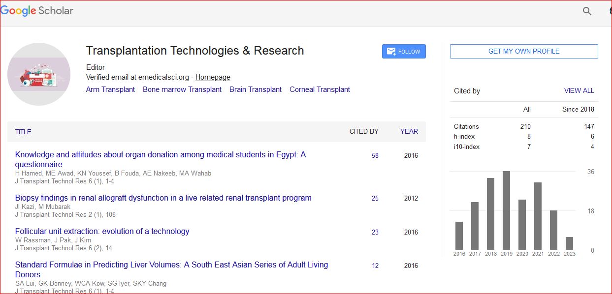
Transplantation Technologies & Research received 223 citations as per Google Scholar report