Zi Qin Ng, Bulang He, Lingjun Mou, Luc Delriviere and Jeffrey M Hamdorf
DOI: 10.4172/2161-0991.1000156
Aim: Various ureteroneocystostomy techniques for kidney transplant have been described with Lich-Gregoir (LG) being widely employed. However, even with multiple modifications on this technique, urine leakage and ureteric stenosis remain as most common complications. This study aims to evaluate urological complications by using our modified LG technique after kidney transplant.
Method: From 26th January 2010 to 30th May 2014, 206 consecutive kidney transplants were performed at our institute. 124 were deceased-donor and 69 were live-donor kidney transplants; 13 patients received a small tumour excised kidney graft. All transplants except one were done by conventional open surgery. The modification involves an additional stitch placed at proximal part of bladder muscular incision with peri ureteric tissue at the entrance of ureter to bladder. Urological complications were defined as urine leakage or ureteral stricture. The patients were followed-up from 12 to 64 months.
Results: There was no urine leakage in this cohort. One case of ipsilateral dual-kidney transplant developed distal ureteral stricture secondary to a lymphocele that was treated by laparoscopic fenestration. Subsequently, surgical reconstruction of urinary tract was required and success. Seven cases had mild to moderate hydronephrosis identified on CDU; 4 were due to a lymphocele; 3 were secondary to urinary tract stones. Four patients had renal pelvis prominence on CDU, which was spontaneously resolved with satisfactory renal function.
Conclusion: From this study, it is demonstrated that urine leakage can be prevented and ureteric stricture may be minimised by using this modified LG technique. The ureter was always shortened in an adequate length and a ureteric stent was inserted. This modification is simple and reproducible by placing additional two stitches, which secure the potential gap at the entrance of ureter to the bladder.
Shigehito Miyagi, Kenji Shimizu, Koji Miyazawa, Yuta Kakizaki, Atsushi Fujio, Yasuyuki Hara, Chikashi Nakanishi, Hitoshi Goto, Takashi Kamei, Naoki Kawagishi, Noriaki Ohuchi and Susumu Satomi
DOI: 10.4172/2161-0991.1000157
Objectives: In living-donor liver transplantation (LDLT), microsurgical reconstruction of a hepatic artery is essential but requires challenging techniques, because graft arteries are short and usable vessel grafts are limited. Furthermore, hepatic artery thrombosis can be a lethal complication. Extra-anatomical jump graft reconstruction using free grafts is reported to have a high reocclusion rate. However, this technique is necessary when there is no other option. We report 4 cases of LDLT that required extra-anatomical reconstruction technique using free autografting from the aorta to the hepatic artery. In this technique, we used the systemic administration of gabexate mesilate that is the strong serine protease inhibitor.
Methods: From 1991 to 2015, we performed 164 LDLTs. We retrospectively investigated 4 cases of extraanatomical reconstruction of the hepatic artery using free autografting from the aorta to the hepatic artery.
Results: Two cases initially underwent anatomical reconstruction, but the arteries occluded early, because of the dissection of recipient’s artery. There was no arterial graft, so we performed extra-anatomical reconstruction by using free autografting from the aorta to the hepatic artery. In the other two cases, the recipient arteries could not be used. Therefore, we initially performed extra-anatomical reconstruction by using radial artery free autografting as jump grafts from the aorta to the hepatic artery. In all cases, we used the systemic administration of gabexate mesilate, and could rescue all cases.
Conclusion: We experienced and were able to salvage 4 cases that required free autografts. When there is no other means of reconstructing arteries, it is necessary to perform this procedure, depending on the condition of the intima of the recipient artery.
William Rassman, Jae Pak and Jino Kim
DOI: 10.4172/2161-0991.1000158
Follicular Unit Extraction (FUE) hair transplantation began as a clinical offering in 2002. Since that time, this minimally invasive hair transplant surgery has grown to a market size of approximately $1.2 billion annually (48.5% of the total hair transplant business world-wide) and is continuing to grow rapidly. This growth is driven by a rapid expansion of the provider pool. New doctors, previous not in the business, have been entering the field and bringing with them, new patients from their own patient populations. The problems that they are encountering are similar to the historic challenges which are outlined in this article updated by the newer instrumentation that has evolved since 2002. Service organizations have arisen where non-professionals are performing the surgery for physicians unable to do so. This article summarizes the evolution of the FUE technology, which has not followed traditional new technological surgical procedures for training new doctors.
Physician innovation became critical in the dissemination of FUE and many doctors previously in the field have had difficulty keeping up. The idea of a minimally invasive FUE technology seems to take on a favorable ‘aire’ for potential patients and for those who heretofore would never have considered having a hair transplant is now coming forward. The authors believe that significant continued changes in the technology are an inevitable outcome of both the rise in the provider pool and the demand for these services. FUE has changed the labor pool as well. The authors have tried to outline the technical changes that impact both labor and the delivery of a better quality outcome provided that the doctors who rally to this opportunity get the proper training that they require. Proper training, unfortunately, seems to have taken a back seat as the financial incentives for the physician has put the cart before the horse.
Fakhriya Alalawi, Hind AlNour, Bridson M Julie, Ajay Sharma and Ahmed Halawa
DOI: 10.4172/2161-0991.1000159
Intra-abdominal and biliary infections are significant cause of morbidity and mortality for the transplant recipients, specifically in the early post-operative period, reflecting substantial immunosuppression. In renal transplant recipients, there is no reported increased risk for pyogenic liver abscess as compared to general population. The diagnosis of hepatic abscess in a transplant recipient can be challenging since presentation could be atypical and the signs and symptoms might be insidious and subtle. If a hepatic abscess is missed and left untreated, it is potentially lethal; hence, an early diagnosis and timely initiation of appropriate therapy is of utmost importance, to improve patient outcomes.
We present a case record of a 29 year old male patient who received a living unrelated renal transplant in 2007. He presented 8 years post-transplant with a fever of unknown origin; clinical and laboratory tests did not provide any clue to the diagnosis. However, labeled white cell scan (Leukoscan) followed by abdominal ultrasound scan (USS) revealed an unexpected finding of a large liver abscess (5.57 × 6.05 cm). He responded well to a course of antibiotics including metronidazole therapy besides requiring percutaneous abscess drainage.
Sergio Brasil, Marcelo de L Oliveira, Manoel J Teixeira, Luiz Marcelo S Malbouisson and Edson Bor-Seng-Shu
DOI: 10.4172/2161-0991.1000160
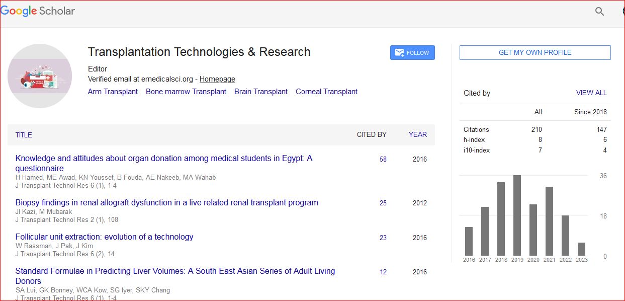
Transplantation Technologies & Research received 223 citations as per Google Scholar report

 Spanish
  Chinese
  Russian
  German
  French
  Japanese
  Portuguese
  Hindi
Hindi 