Andrew J Kobets, Adam Ammar, Rick Abbott and James T Goodrich
Children's Hospital at Montefiore, USA
Scientific Tracks Abstracts: J Material Sci Eng
Objective: Sagittal synostosis affects 1 in 1000 live births and may result in increased intracranial pressure, hindrance
of normal neural development, and cosmetic deformity due to scaphocephaly. Historically, several approaches have
been utilized for surgical correction and recently, computed tomography (CT)-guided reconstruction procedures
are increasingly used. In this report, the authors describe the use of a CT-derived virtual and stereolithographic
(3D printed) craniofacial models, which are used to guide intraoperative bone placement, and intraoperative CT
guidance for confirmation of bone placement, to ensure the accuracy of surgical correction of scaphocephaly, as
demonstrated to parents.
Methods:Preoperative high-resolution CT imaging was used to construct 3D image sets of the skulls of two infants
(a 14-month-old female and a 6-month-old male) with scaphocephaly. These 3D image sets were then used to create
a virtual model of the proposed surgical correction for each of the infants�?? deformities, which was then printed and
made available for use intra-operatively to plan the bone flap, fashion the bone cuts, and optimize graft placement.
After the remodeling, adherence to the preoperative plan was assessed by overlaying a CT scan of the remodeled
skull with the virtual model. Deviations from the preoperative model were noted.
Findings: Both patients had excellent postoperative cosmetic correction of head shape and contouring. The mean
operative time was 5 h, blood loss was 100 ml, and one child required modification of the subocciput after intraoperative
imaging showed a deviation of the reconstruction from the surgical goal as depicted by the preoperative model.
Conclusion: The addition of neuro-navigation to stereolithographic modeling ensured the accuracy of the
reconstruction for our patients and provided greater confidence to both surgeons and parents. While unisutural cases
are presented for clarity, correction was still required for one patient. The cost of the models and the additional CT
required must be weighed against the complexity of the procedure and possibly reserved for patients with potentially
complicated corrections.
Recent Publication:
1. 1. Alvarez Garijo J A, Cavadas P C, Vila M M and Alvarez Llanas A (2001) Sagittal synostosis: results of surgical
treatment in 210 patients. Childs Nerv Syst 17:64-68.
2. 2. Anderson P J, Yong R, Surman T L, Rajion Z A and Ranjitkar S (2014) Application of three-dimensional
computed tomography in craniofacial clinical practice and research. Aust Dent J 59(Suppl 1):174-185.
3. 3. Bendon C L, Johnson H P, Judge A D, Wall S A and Johnson D (2014) The aesthetic outcome of surgical
correction for sagittal synostosis can be reliably scored by a novel method of preoperative and postoperative
visual assessment. Plast Reconstr Surg 134:775e-786e.
4. 4. Bly R A, Chang S, Cudejkova M, Liu J J and Moe K S (2013) Computer-guided orbital reconstruction to
improve outcomes. JAMA Facial Plastic Surgery 15:113-120.
5. 5. Darwood A, Collier J, Joshi N, Grant W E, Sauret Jackson V, Richards R, Dawood A and Kirkpatrick N
(2015) Re-thinking 3D printing: a novel approach to guided facial contouring. J Craniomaxillofac Surg
43(7):1256-1260.
Andrew J Kobets is currently Chief Resident of Neurosurgical Resident at Montefiore Medical Center and is working on a translational research project as a Visiting Scientist at both the Feinstein Institute for Medical Research at Northwell Health and at the Albert Einstein College of Medicine. He has initiated and overseen the initiation of three clinical trials in the Department of Neurological Surgery at Montefiore Medical Center and is the first utilizing MR Elastography to evaluation shunt function in New York. He graduated from the Yale School of Medicine with a medical degree and a Master’s in Health Sciences. He studied Systems Neuroscience at the Johns Hopkins University as an Undergraduate and will return to Johns Hopkins after residency for a Fellowship in Pediatric Neurosurgery.
E-mail: akobets@montefiore.org
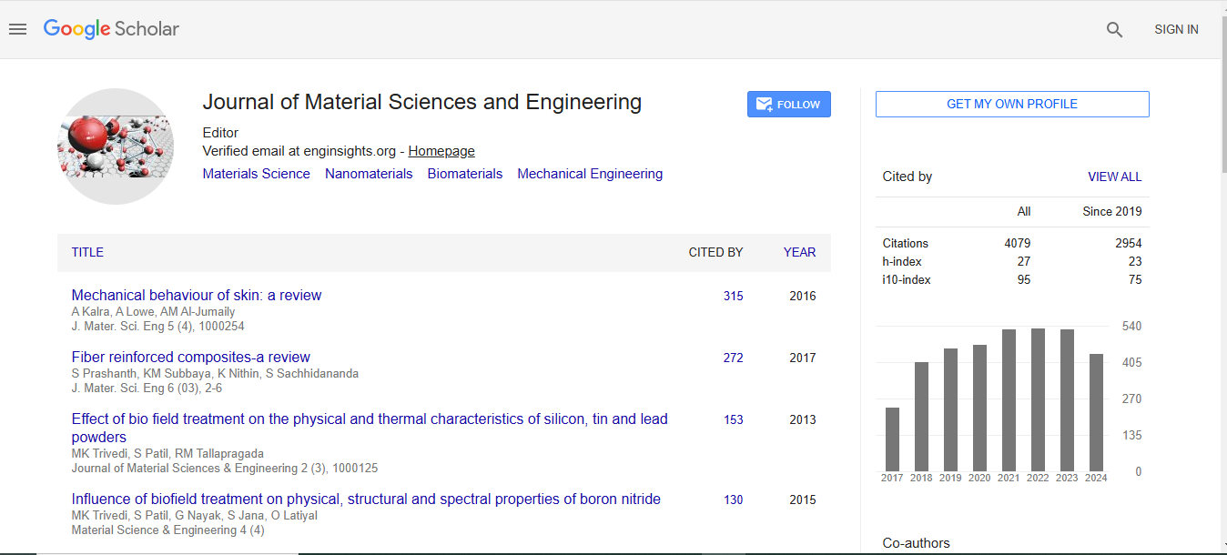
Journal of Material Sciences & Engineering received 3677 citations as per Google Scholar report

 Spanish
  Chinese
  Russian
  German
  French
  Japanese
  Portuguese
  Hindi
Hindi 