Dimitar Stamov, Sandra Kostrowski, Torsten J�?¤hnke, and Heiko Haschke
JPK Instruments, Germany
Scientific Tracks Abstracts: J Material Sci Eng
Besides structural and physico-chemical composition, the topography, roughness, adhesiveness and mechanical properties of biomaterials are relevant parameters which strongly affect cell differentiation and tissue formation, and are thus crucial for assessing biocompatibility in the human body. We have developed a multipurpose AFM device which allows comprehensive characterization of these properties and interactions on the nanoscale under physiological conditions and in combination with advanced optical microscopy. Our unique quantitative imaging (QIâ�?¢) mode determines several sample properties, like topography and the Youngâ�?�?s modulus, with one measurement. With the CellHesion�?® technique, the adhesion of a single cell to any substrate can be measured and validated. The NanoWizard�?® ULTRA Speed technique enables fast AFM imaging of dynamic processes with approx. 1 frame per second. Using QIâ�?¢, we have characterized the topography and mechanical properties of challenging samples like living cells and tissue sections. The adhesion of single fibroblast cells to various surface modifications could be quantified and their suitability as cochlear implant coatings could be assessed. Fast AFM imaging revealed collagen type I fibrillogenesis and the formation of the 67 nm D-banding in situ with high spatio-temporal resolution. Here, we are presenting an enhanced AFM, making this technique a valuable tool for biomedical research.
Dimitar Stamov received his Master of Science (M.Sc.) degree in Molecular Bioengineering from the Technical University of Dresden, Germany in 2005. He continued with his gradual studies at the Leibniz Institute of Polymer Research Dresden (with TU Dresden) in the field of structural analysis of extracellular matrix assemblies and proteins. In 2011 he started post-doctoral work in the NanoBiology group at the DFG-Center for Functional Nanostructures at the Karlsruhe Institute of Technology (KIT). Since 2013 he joined JPK Instruments AG, a manufacturer of nano-analytical instruments based on atomic force microscopes and optical tweezers, as an application scientist. He is currently focusing on the application of high-resolution and fast scanning techniques for the full characterisation of life science, soft matter, as well as dynamic systems.
Email: stamov@jpk.com
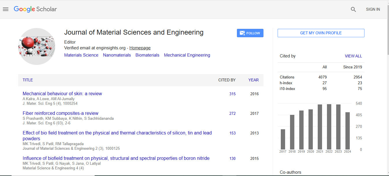
Journal of Material Sciences & Engineering received 3677 citations as per Google Scholar report

 Spanish
  Chinese
  Russian
  German
  French
  Japanese
  Portuguese
  Hindi
Hindi 