Mini Review - (2023) Volume 14, Issue 1
Received: 30-Jan-2023, Manuscript No. jbsbe-23-102167;
Editor assigned: 01-Feb-2023, Pre QC No. P- 102167;
Reviewed: 13-Feb-2023, QC No. Q- 102167;
Revised: 21-Feb-2023, Manuscript No. R- 102167;
Published:
27-Feb-2023
, DOI: 10.37421/2155-6210.2023.14.374
Citation: Lubica, Luiliana. “Utilizing Nanopore Sensing Techniques for the Detection of Biological Molecules.” J Biosens Bioelectron 14 (2023): 374.
Copyright: © 2023 Lubica L. This is an open-access article distributed under the terms of the Creative Commons Attribution License, which permits unrestricted use, distribution, and reproduction in any medium, provided the original author and source are credited.
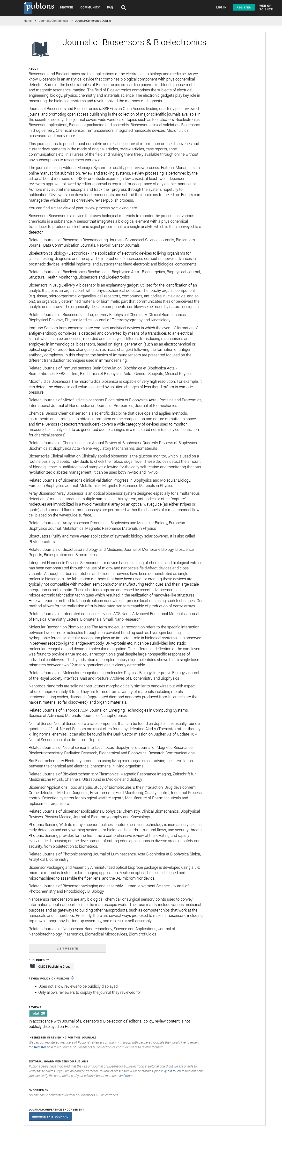
Present day biomedical detecting procedures have fundamentally expanded in accuracy and exactness because of new innovations that empower speed and that can be custom fitted to be profoundly unambiguous for markers of a specific sickness. Diagnosing beginning phase conditions is vital to treating serious sicknesses. Generally, in the beginning phases of the sickness, the quantity of explicit biomarkers is extremely low and at times challenging to distinguish utilizing traditional analytic strategies. Biosensors are currently generating a lot of interest in the medical field due to their ease of use, portability, and speed, as well as their low costs and consistent, dependable results. The ability of low-concentration single-molecule sensors like nanopores to detect biomolecules has the potential to become clinically useful. As a result, blood markers, nucleic acids, and protein detection applications have emerged in this field. The utilization of nanopores presently can't seem to arrive at development for normalization as analytic methods, not withstanding, they guarantee huge potential, as headway is made into settling nanopore structures, upgrading sciences, and further developing information assortment and bioinformatic examination. Based on various types of nanopores, this review provides a fresh perspective on current biomolecule sensing methods, obstacles, and strategies for clinical application.
Diagnostics • Biosensors • Clinical applications • Ionic currents • Polymers • Nanopores
As a result of ongoing lifestyle changes and advancements in our knowledge of the human body, new health conditions and the mechanisms that underlie them are discovered every year. The identification of particular molecules involved is crucial for human health due to the intrinsic molecular basis of disease causes and manifestations. Alzheimer's disease, Parkinson's disease, and cancer are all diseases that would benefit from early detection because they can significantly shorten life expectancy and quality of life. A fascinating and significant methodology toward this path is the discovery of biomarkers at the single particle level through nanopores. Structures known as nanopores can either be made from synthetic materials like nanoscale silicon or graphene or can occur naturally as proteic polymers. Due to changes in the local microenvironment, a nanopore embedded in a dielectric membrane can be used to detect biomolecules, particularly DNA, RNA, and proteins. From the discovery of the α-HL (α-hemolysin) organization in lipidic solutions to solid nanopores based on silicon, graphene, etc., over time, various organic and synthetic nanopores, have been utilized in molecular biology to generate consistent information. Due to their adaptable geometry and shapes, solid nanopores are widely used and can be produced depending on the analyte being detected. As a result, the solid-state nanopore only performs a few detections with a low degree of selectivity because it does not undergo selective translocations. Late examinations have researched this issue and functionalized stable surfaces with acknowledgment particles that permit them to distinguish a particular sub-atomic element. When an electrical potential is applied across a nanopore, it creates a current. When a molecule enters the nanopores, they either completely or partially block the flow. This blockage is described by the adjustment of flow and stay time, which relates to the size and separate electric charge of the particle [1,2].
The physical measurement of the net transport of charged loads per unit of time is the current that enters a nanopore. Accordingly, the species entering the nanopore may impact the adequacy of the current, which is firmly connected with the properties of the analyte, nanopore, and arrangement The properties of the translocating analyte, hydrodynamic effects (electro-osmosis), molecular interactions, the surface of the local charge distribution (polarization, concentration of condensation ions), and the type of molecules that cross the nanopore are all crucial. Changing the properties of strong nanopores may change movement time, which is the time it takes for the particle to go through the nanopore from one side to the next, free of other exploratory boundaries [3]. For instance, the dwell time, or the amount of time a molecule spends inside the analyte's nanopore, can be significantly altered by even the smallest change in the superficial load. The electrophysiological solution (electrolyte), the physicochemical properties of the molecule of interest (analyte), and the potential applied from the outside all have an impact on how a molecule moves through a nanopore The objectives of this review are to both emphasize the adaptability of the nanopore analysis technique and shed light on the difficulties it poses in relation to the molecule under investigation. The paper contains a number of distinct experimental aspects for various molecules that are significant for human health [4].
Because of their biosensing abilities, nanopores are the reason for the advancement of new advancements for the fast recognition of different explicit particles engaged with a few pathologies. Numerous studies have emphasized the significance of utilizing nanopores. Using nanopores, genetic analyses, for instance, can be completed in as little as seven hours, providing patients with a definitive diagnosis in a short amount of time [whereas conventional methods typically require several days or even weeks. In addition, the sequence, structure, and components of proteins in living organisms contain information that is crucial to physiological processes, whether normal or abnormal, but numerous obstacles and deficiencies remain in this area. For instance, it is difficult to identify signal fluctuations that ought to be unique to the translocated molecule if molecules are moved through the nanopore too rapidly. Because of this, researchers looked into ways to slow down the transport of molecules. The average duration and amplitude of current blockages typically contain information about an analyte's identity, but there are numerous strategies to improve nanopore analysis methods by adjusting various experimental parameters to achieve high resolution. A distinct charge profile, a three-dimensional structure, and an amino acid sequence characterize each protein. When passing through a nanopore, this information is reflected in the detected current signal. This principle has been used to investigate various properties of proteins or other molecules of interest in the nanopore domain. Nanopores are also utilized in accordance with the properties of the molecules of interest [5,6].
It's possible that utilizing nanopores as a method of analysis for the purpose of diagnosing a variety of diseases is a novel approach that holds promise to go far beyond the analyses that are currently carried out in hospitals. The nanopore assay method promises to revolutionize both traditional biomarker molecule analysis and molecular detection at low concentrations and high resolution. The technology's use at the point of care has enormous potential because it is now so small. Real-time analysis, which permits the dynamic monitoring of biological processes, is one of the most significant benefits of the nanopore analysis method. However, this method is not yet widely used due to a number of obstacles. In addition to the physical and chemical optimizations that are necessary for each kind of nanopore, including the structure and materials of the nanopores-biological or synthetic, there are numerous difficulties with molecule capture, controlling the translocation rate, and preventing blockages caused by the analysis of molecules that are too big or interact with the pore surface. Attention to both the sample and the experimental parameters, such as applied voltage, buffer pH, external noise, and solution purity, is necessary for better resolution in nanopore experiments. In the case of OmpG nanopores, for instance, the pH of the electrophysiological solution can have an effect on them at an acidic pH, they can either close or influence transport processes through the nanopore.
The method is currently being refined in terms of both working time and outcomes. When compared to culture media, for instance, results can be obtained in as little as six hours. The bioinformatics analysis of the signal is another factor that has an impact on the outcomes. When DNA sequencing uses the base-calling method to determine the DNA structure, bioinformatics refers to the analysis and interpretation of the resulting electrical signal to arrive at biologically or medically conclusive results. Algorithms that make an effort to analyze the signal make use of cutting-edge techniques like machine learning, Markov models, and neural networks. Time-to-value ratios are significantly improved thanks to the development of nanopore analysis techniques made possible by all of these factors.
None.
There are no conflicts of interest by author.
Google Scholar, Crossref, Indexed at
Google Scholar, Crossref, Indexed at
Google Scholar, Crossref, Indexed at
Google Scholar, Crossref, Indexed at
Google Scholar, Crossref, Indexed at
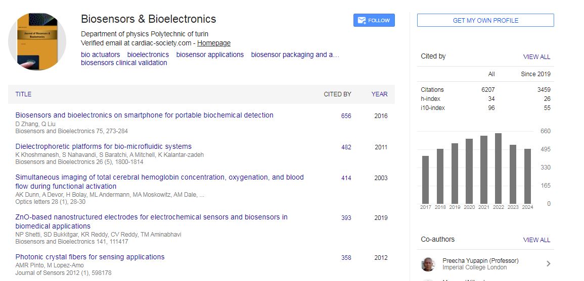
Biosensors & Bioelectronics received 6207 citations as per Google Scholar report