Commentary - (2023) Volume 7, Issue 3
Received: 01-May-2023, Manuscript No. hps-23-105843;
Editor assigned: 03-May-2023, Pre QC No. P-105843;
Reviewed: 15-May-2023, QC No. Q-105843;
Revised: 20-May-2023, Manuscript No. R-105843;
Published:
27-May-2023
, DOI: 10.37421/2573-4563.2023.7.222
Citation: Oquendo, Dorothea. “Unraveling the Matrix:
Understanding Liver Fibrosis and its Extracellular Matrix Deposition.” J Hepatol
Pancreat Sci 7(2023): 222.
Copyright: © 2023 Oquendo D. This is an open-access article distributed under the terms of the Creative Commons Attribution License, which permits unrestricted use, distribution, and reproduction in any medium, provided the original author and source are credited.
Hepatic Stellate Cells (HSCs), also known as per sinusoidal cells or Ito cells, are a specialized population of cells residing in the livers per sinusoidal space, also known as the space of Disse. HSCs play a crucial role in the normal liver function, as well as in the pathogenesis of liver fibrosis. These cells are quiescent in healthy liver tissue but become activated in response to liver injury, triggering a cascade of cellular and molecular events leading to fibrosis. In their quiescent state, HSCs are characterized by their location within the space of Disse and their unique morphology. Quiescent HSCs contain lipid droplets rich in vitamin A, giving them a characteristic star-shaped appearance when viewed under a microscope. They are responsible for storing and releasing vitamin A, which is essential for the liver's metabolic functions. Upon liver injury, HSCs undergo a phenotypic transformation into activated my fibroblasts, which are the primary producers of Extracellular Matrix (ECM) proteins during liver fibrosis. Activated HSCs lose their lipid droplets and acquire contractile properties, allowing them to migrate and proliferate within the liver tissue. This activation process is triggered by various profibrotic stimuli, such as cytokines, growth factors, and oxidative stress [1]. Activated HSCs contribute to liver fibrosis through their ability to synthesize and secrete excessive amounts of ECM proteins, including collagen, fibronectin, and elastin. This leads to the formation of fibrotic scar tissue, which replaces the normal liver parenchyma and disrupts the organ's structure and function. The activated HSCs also play a role in inflammation by producing pro-inflammatory cytokines and chemokines, further exacerbating the fibrotic process. Transforming Growth Factor-beta (TGF-β) is a key cytokine involved in HSC activation, promoting their transition from a quiescent to an activated state. Other signalling pathways, such as the Platelet-Derived Growth Factor (PDGF), hedgehog, and Wnt/β-catenin pathways, also contribute to HSC activation and ECM synthesis.
Kupffer cells, the resident macrophages of the liver, play a crucial role in maintaining liver homeostasis and immune surveillance. These specialized cells are strategically located within the sinusoidal lining of the liver, where they constantly monitor the blood for pathogens, toxins, and other foreign substances [2]. When the liver encounters injury or infection, Kupffer cells rapidly respond by initiating an inflammatory response. They possess Pattern Recognition Receptors (PRRs), such as Toll-Like Receptors (TLRs), which enable them to recognize and respond to Pathogen-Associated Molecular Patterns (PAMPs) and Danger-Associated Molecular Patterns (DAMPs). Upon activation, Kupffer cells release a variety of pro-inflammatory cytokines, chemokines, and Reactive Oxygen Species (ROS) to recruit and activate other immune cells.
The activation of Kupffer cells in response to liver injury is a complex process involving various signalling pathways and molecular mediators. The release of pro-inflammatory cytokines, such as Tumor Necrosis Factor-alpha (TNF-α), Interleukin-1 beta (IL-1β), and Interleukin-6 (IL-6), by activated Kupffer cells contributes to the recruitment and activation of other immune cells, such as neutrophils and monocytes [3]. The sustained activation of Kupffer cells in chronic liver diseases leads to the production of pro-fibrotic mediators, including transforming growth factor-beta (TGF-β), Platelet-Derived Growth Factor (PDGF), and Connective Tissue Growth Factor (CTGF). These factors promote the activation of Hepatic Stellate Cells (HSCs) - the main ECM-producing cells in liver fibrosis - and drive the progression of fibrosis.
Epithelial-Mesenchymal Transition (EMT) is a biological process that involves the conversion of epithelial cells into mesenchymal cells. It plays a crucial role in various physiological and pathological processes, including embryogenesis, tissue repair, cancer metastasis, and organ fibrosis, including liver fibrosis. In EMT, epithelial cells lose their characteristic cell-cell adhesion and acquire mesenchymal-like characteristics, such as enhanced migratory capacity, invasive behaviour, and increased production of Extracellular Matrix (ECM) proteins. This phenotypic transformation is regulated by a complex network of signalling pathways and transcription factors. During EMT, the epithelial cells undergo several characteristic changes. The cells lose their apical-basal polarity and cell-cell junctions, such as tight junctions and adherens junctions, which are critical for maintaining epithelial integrity. This disruption of cell-cell adhesion allows the cells to detach from the epithelial layer and acquire a more motile mesenchymal phenotype. One of the key features of EMT is the down regulation of epithelial markers, such as E-cadherin and cytokeratin’s, and the up regulation of mesenchymal markers, including N-cadherin, vimentin, and fibronectin.
Liver fibrosis is a dynamic and progressive process that develops as a result of chronic liver injury. It involves a complex interplay of cellular and molecular events, leading to the excessive deposition of Extracellular Matrix (ECM) proteins and the disruption of normal liver architecture. Understanding the pathogenesis and progression of liver fibrosis is crucial for developing effective therapeutic strategies to prevent or reverse the fibrotic process. The initiation of liver fibrosis begins with the underlying cause of chronic liver injury. This can be due to various etiologies, such as Viral Hepatitis (B and C), alcohol abuse, Non-Alcoholic Fatty Liver Disease (NAFLD), autoimmune liver diseases, or drug-induced liver injury. Hepatocyte injury, inflammation, and oxidative stress are common triggers that initiate the fibrogenic response. The inflammatory phase of liver fibrosis is characterized by the activation of immune cells, such as Kupffer cells and infiltrating inflammatory cells, including neutrophils and monocytes. These cells release pro-inflammatory cytokines, chemokines, and growth factors that promote hepatocyte damage and recruit additional immune cells to the site of injury.
Accurate diagnosis and staging of liver fibrosis are crucial for guiding treatment decisions, monitoring disease progression, and evaluating therapeutic efficacy. Transient Elastography (TE) is a widely used non-invasive technique that measures liver stiffness as an indirect marker of fibrosis. TE utilizes ultrasound to assess liver stiffness, providing a quantitative measure of fibrosis severity. Other imaging techniques, such as Magnetic Resonance Elastography (MRE), Shear Wave Elastography (SWE), and Acoustic Radiation Force Impulse Imaging (ARFI), also measure liver stiffness and have shown promising results in fibrosis assessment.
Several serum biomarkers include direct markers of fibrogenesis, such as Hyaluronic Acid (HA), Procollagen type III Peptide (PIIINP), and Amino-terminal Propeptide of type III Collagen (PIIINP), as well as indirect markers of fibrosis, such as Fibrosis-4 Index (FIB-4), Aspartate Aminotransferase-to-Platelet Ratio Index (APRI), and Enhanced Liver Fibrosis (ELF) score. These biomarkers are often used in combination or as part of panels to enhance diagnostic accuracy. Imaging-based techniques, such as Magnetic Resonance Imaging (MRI) and Computed Tomography (CT) scans, can provide valuable information about liver morphology, fibrotic changes, and associated complications [4].
Liver fibrosis is a progressive condition that can lead to serious complications, such as cirrhosis and liver failure. Effective therapeutic strategies for liver fibrosis aim to halt or reverse the fibrotic process, promote liver regeneration, and restore normal liver function. ECM modulation aims to promote the remodelling and resolution of fibrotic scar tissue. This approach involves targeting enzymes involved in ECM turnover, such as matrix Metalloproteinase (MMPs) and Tissue Inhibitors of Metalloproteinase (TIMPs), to restore the balance between ECM synthesis and degradation. Therapies targeting specific ECM components, such as collagen cross-linking inhibitors, are also being investigated. Inflammation plays a critical role in the pathogenesis of liver fibrosis [5]. Therapies aimed at reducing inflammation and modulating the immune response can potentially slow down fibrotic progression. Anti-inflammatory drugs, immunomodulatory agents, and immune checkpoint inhibitors are being explored as potential therapeutic options.
Metabolic interventions focus on modifying underlying metabolic abnormalities that contribute to liver fibrosis, particularly in cases of Non- Alcoholic Fatty Liver Disease (NAFLD) and Non-Alcoholic Steatohepatitis (NASH). Lifestyle modifications, including weight loss, exercise, and dietary changes, are fundamental in managing these conditions. Additionally, pharmacological interventions targeting metabolic pathways involved in hepatic lipid accumulation and inflammation, such as Peroxisome Proliferator- Activated Receptor (PPAR) agonists, are being investigated.
None.
There are no conflicts of interest by author.
Google Scholar, Crossref, Indexed at
Google Scholar, Crossref, Indexed at
Google Scholar, Crossref, Indexed at
Google Scholar, Crossref, Indexed at
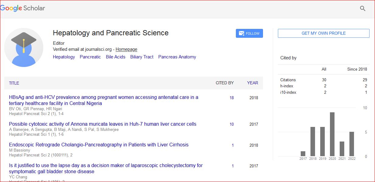
Hepatology and Pancreatic Science received 34 citations as per Google Scholar report