Brief Report - (2023) Volume 9, Issue 1
Received: 31-Jan-2023, Manuscript No. aso-23-98367;
Editor assigned: 02-Feb-2023, Pre QC No. P-98367;
Reviewed: 16-Feb-2023, QC No. Q-98367;
Revised: 21-Feb-2023, Manuscript No. R-98367;
Published:
28-Feb-2023
, DOI: 10.37421/2471-2671.2023.9.35
Citation: Marx, Wilkinson. "Surgical Approaches for Biliary Rhabdomyosarcoma: Laparotomies as Preferred Treatment Option.’’ Arch Surg Oncol 9 (2023): 35.
Copyright: © 2023 Marx W. This is an open-access article distributed under the terms of the Creative Commons Attribution License, which permits unrestricted use, distribution, and reproduction in any medium, provided the original author and source are credited.
Rhabdomyosarcoma (RMS) is a rare form of cancer that affects children. It is a malignant tumor that develops in the muscles that are responsible for movement, such as those in the arms, legs and torso. RMS can occur in any part of the body, but it is most commonly found in the head and neck area, followed by the genitourinary tract, extremities and trunk. RMS is a type of soft tissue sarcoma, which means it originates in the body's connective tissues, including muscle, fat and fibrous tissue. RMS is the most common type of soft tissue sarcoma in children, with around 350 new cases diagnosed in the United States each year.
Biliary rhabdomyosarcoma • Laparotomies • Treatment
The exact cause of RMS is not known, but it is believed to be caused by changes (mutations) in the DNA of developing muscle cells. Some genetic syndromes, such as Li-Fraumeni syndrome and neurofibromatosis, may increase the risk of developing RMS. The symptoms of RMS can vary depending on the location of the tumor. In some cases, there may be no symptoms at all, while in others, the tumor may cause pain, swelling, or a noticeable lump. Other symptoms may include difficulty breathing, difficulty swallowing, or changes in bowel or bladder function.
Diagnosis of RMS typically involves a physical exam, imaging tests (such as X-rays, CT scans, or MRIs) and a biopsy to confirm the presence of cancerous cells. Once RMS is diagnosed, treatment typically involves a combination of surgery, radiation therapy and chemotherapy. The specific treatment plan will depend on the location, size and stage of the tumor, as well as the child's overall health. The prognosis for RMS varies depending on the stage of the tumor, the location of the tumor and other factors. In general, children with localized tumors have a better prognosis than those with metastatic tumors that have spread to other parts of the body. With early diagnosis and appropriate treatment, many children with RMS can achieve long-term remission and lead normal, healthy lives [1].
RMS is a rare malignant tumor affecting children that develops in the muscles responsible for movement. Its cause is not fully understood and symptoms vary depending on the location of the tumor. Early diagnosis and appropriate treatment can lead to long-term remission and a good prognosis for children with RMS. Ongoing research into the causes and treatment of RMS is critical to improving outcomes for children with this rare cancer.
Rhabdomyosarcoma (RMS) is a rare form of cancer that primarily affects children. It develops in the muscles that are responsible for movement and can occur in any part of the body, including the digestive system. One area where RMS is worth considering when dealing with medical conditions is incase of choledochal cysts. A choledochal cyst is a rare congenital malformation of the bile ducts that affects both adults and children. It is characterized by the dilation or expansion of the bile ducts that carry bile from the liver to the small intestine. The exact cause of choledochal cysts is not known, but they are thought to be related to a congenital defect that occurs during fetal development [2-4].
While choledochal cysts themselves are not cancerous, they do have an increased risk of developing cancerous growths. In particular, choledochal cysts are associated with an increased risk of developing bile duct cancer, which is a rare but aggressive form of cancer. Recent research has shown that there is a significant association between RMS and choledochal cysts. Studies have found that children with choledochal cysts are at increased risk of developing RMS, particularly in the presence of biliary dilatation or obstruction.
One possible explanation for this link is that the abnormal development of the bile ducts that leads to choledochal cysts may also create an environment that promotes the development of RMS. Alternatively, the prolonged exposure to inflammation and infection associated with choledochal cysts may increase the risk of developing RMS. It is important for medical professionals to consider the possibility of RMS when dealing with choledochal cysts, particularly in cases where there is biliary obstruction or dilatation. Early diagnosis and appropriate treatment are critical for achieving the best possible outcomes for children with RMS.
While RMS is a rare form of cancer that primarily affects children, recent research has shown that it is worth considering when dealing with choledochal cysts. Children with choledochal cysts are at an increased risk of developing RMS, particularly in the presence of biliary dilatation or obstruction. Medical professionals should be aware of this link and consider the possibility of RMS when dealing with choledochal cysts, as early diagnosis and appropriate treatment are critical for achieving the best possible outcomes. Rhabdomyosarcoma (RMS) is a rare type of cancer that primarily affects children and adolescents. It arises from the muscle tissue and can occur in various parts of the body, including the biliary system. The biliary system comprises the liver, gallbladder and bile ducts, which are responsible for producing and transporting bile to the small intestine. When RMS occurs in the biliary system, surgical intervention is often required.
Laparotomy is a surgical procedure that involves making a large incision in the abdomen to gain access to the abdominal organs. This approach allows surgeons to visualize and manipulate the organs directly, making it an effective technique for treating various abdominal pathologies, including biliary RMS. The surgical approach for biliary RMS depends on several factors, such as the location and extent of the tumor, the age and health status of the patient and the expertise and experience of the surgical team. The primary aim of surgery for biliary RMS is to remove the tumor completely while preserving the function of the biliary system and minimizing postoperative complications.
Several surgical approaches have been reported for the management of biliary RMS, including laparoscopic and open procedures. However, most studies report the use of laparotomy for the treatment of biliary RMS. Laparotomy is preferred for larger tumors, tumors located in difficult-to-reach areas and cases with suspected involvement of adjacent organs. During a laparotomy for biliary RMS, the surgeon makes an incision in the abdomen to expose the affected organs. The tumor is carefully dissected from the surrounding tissues and removed completely, along with any involved lymph nodes. If necessary, the surgeon may also perform a bile duct resection or reconstruction to restore the normal flow of bile [5].
Following the surgical removal of the tumor, additional treatments, such as chemotherapy and radiation therapy, may be recommended to prevent recurrence and improve overall survival. The use of adjuvant therapies is typically decided on a case-by-case basis, depending on the stage and extent of the tumor. Biliary RMS is a rare type of cancer that requires surgical intervention for the best chance of a cure. While laparotomy is not the only surgical approach used for the management of biliary RMS, it is the most commonly reported in the literature. Laparotomy allows for the complete removal of the tumor while minimizing complications and preserving biliary function. Additional therapies may also be required to improve outcomes and reduce the risk of recurrence. Patients diagnosed with biliary RMS should consult with a specialized surgical team to determine the best course of treatment for their individual case.
None.
None.
Google Scholar, Crossref, Indexed at
Google Scholar, Crossref, Indexed at
Google Scholar, Crossref, Indexed at
Google Scholar, Crossref, Indexed at
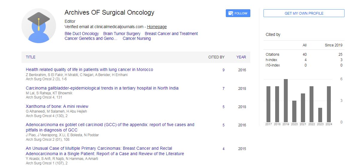
Archives of Surgical Oncology received 37 citations as per Google Scholar report