Research Article - (2022) Volume 13, Issue 3
Received: 07-Jul-2022, Manuscript No. JTESE-22-68880;
Editor assigned: 11-Jul-2022, Pre QC No. JTESE-22-68880(PQ);
Reviewed: 26-Jul-2022, QC No. JTESE-22-68880;
Revised: 06-Sep-2022, Manuscript No. JTESE-22-68880(R);
Published:
14-Sep-2022
, DOI: 10.37421/2165-8064.2022.12.501
Citation: Bansode, Savita H, Vasant Khare Priyanka and Mahanwar PA. "PLGA Copolymer Synthesis and Nanofabrication by Electro and Melt Spinning." J Textile Sci Eng 12 (2022): 501.
Copyright: © 2022 Bansode SH, et al. This is an open-access article distributed under the terms of the creative commons attribution license which permits
unrestricted use, distribution and reproduction in any medium, provided the original author and source are credited.
Polylactic Glycolic Acid (PLGA) is most important polymer in biomedical applications because we can modify the degradation rate by copolymerization ratio, processing. The ideal scaffold should be three dimensional, highly porous, biodegradable and biocompatible without immune reaction or inflammation. In addition, it should have proper mechanical properties to support the growth of new tissue. Increased in the use of electrospinning nanofiber technique to create nanofiber scaffold for tissue engineering, as there are reports that these scaffolds successfully promote to cell matrix and cell-cell interactions with the cells of human body. Now days, success have been achieved in skin, bladder, airway, bone, kidney where tissue engineering construct has been successfully used.
PLGA synthesis done by convectional method, with study of various parameter such as time, temperature, monomer and catalyst ration. PLGA can be synthesized by polycondensation (convectional) method at 130°C, for 25 hours. Important characteristics such as melting temperature, glass transition temperature, and degradation temperature was determined by DSC and TGA analysis, it was obtained as 168.44°C, 55.76°C and 87.61°C respectively. Chemical structure was studied by FTIR and NMR. These results helped to study the effect of monomer, catalyst on reaction and determining the parameters for melt and electro spinning. Because of good biocompatibility and biodegradability, they can be used in various areas, such as long-term release systems and the tissue engineering.
Biodegradable • Electrospinning • Melt spinning • Polymerization • Synthesis
Tissue engineering, is the field which applies knowledge of life science and the principles of engineering towards the developing, maintaining or improving damaged tissue functions of human being. Tissue engineering can act as a substitute for the Extra Cellular Matrix (ECM) and provide a substrate for cellular adhesion and organization [1]. Thus, tissue engineering scaffolds have a strong resemblance to the natural ECM, which is comprised of nanometerdiameter protein fibers [2]. There are five classes of biomaterials; metals, ceramics, polymers, synthetic polymers, and composites. In general, natural and synthetic polymers are used to replace skin, tendon, ligament, breast, eye, vascular systems, and facial tissues, there are different methods for nanofabrication: Self-assembly, phase separation, electrospinning and melt blowing. Major properties of biodegradable polymer are: Non-toxic, capable of maintaining good mechanical property till degradation, controlled rates of degradation [3].
Electrospinning is one of the best methods for polymer nanofabrication has been studied in details [4]. In addition to this, an ideal scaffold should possess the characteristics such as biocompatible and biodegradable, highly porous structure, suitable mechanical strength and a proper surface topography. In contrast to metals and ceramic, polymeric materials such as Poly Lactic Acid (PLA), electrospinning has become most popular due its advantages over the other methods. Electrospinning is a simple and economic method which can be implemented for the continuous production of polymer nanofiber [5].

Figure 1. Electrospinning setup.
The electrospinning device consists of three main components: syringe pump, collector and high voltage supply. A schematic representation of electrospinning is presented in (Figure 1) [6].
The method comprises of following steps:
• The polymer of which the nanofibers have to be produced is dissolved in solvents.
• The solution of the dissolved polymer is inserted into a syringe.
• The tip of the syringe needle is connected to a high-voltage power supply.
• Fibers are collected on the appropriate collector.

Figure 2. Set up of melt spinning.
Melt spinning set up has been presented in Figure 2 melt spinning has a simple setup where fibers from a molten polymer can directly be drawn continuously, avoiding the use of solvents. The fibers, temperature of spinning (extruder) and the rpm of take-up roll were kept constant at 110°C and 900 rpm, respectively. The fibers were air cooled before they wind up on the take-up roll [7,8].
Effect of solution properties
Effect of viscosity/solution concentration: A very high solution concentration (>40 wt.%) resulted in drying of the droplets at the tip which thereby prevented further fibers formation. Another study by Cui reports formation of beaded fibers at low solution concentrations observed during the electrospinning of Poly (DL-Lactide) (PDLLA) [9].
Effect of conductivity or charge density: Addition of salts like sodium chloride, sodium phosphate and potassium phosphate results into uniform smaller beadless fibers with diameters ranging from 200 nm to 1000 nm [10,11]. It is concluded that in the electrospinning process the occurrence of beads can be completely prevented with the addition of suitable salts [12].
Effect of polymer molecular weight: The polymer molecular weight has a direct relation with the solution viscosity. It is observed that the solution viscosity increases with increasing molecular weight of polymer. It was also observed that the number of beads and droplets decreased with increasing molecular weight [13].
Effect of dipole moment and dielectric constant, surface tension: When the polymeric solution is subjected to an electric field, higher dielectric constant and dipole moment of solvents gives higher electrical susceptibility which thereby helps to increase the productivity of the solution [14,15]. In addition, dielectric constant of a solvent roughly provides a major of its polarity. Therefore, it is very important to study the effect of these parameters on the electrospinning. It is observed that higher dielectric constant reduced the beads formation and the fiber diameter in the electrospun fiber [16]. Yang studied the effect of surface tensions on the electrospun nanofibers of poly(vinylpyrrolidone) using various solvents such as ethanol, dichloromethane and N, N-Di Methyl Formamide (DMF) [17,18].
PLGA Synthesis (Convectional synthesis)
Materials: Lactic acid 90% pure was purchased from Loba chemie, used as monomer. 97% pure Glycolic acid was purchased from Sigma Aldrich, as monomer. Stannous octoate 99% pure was purchased from Sigma Aldrich and used as initiator and catalyst (it acts as both). Stearyl alcohol and Lauryl alcohol (reagent grade) from SD fine chemicals Pvt. Ltd used as co-catalyst. Silicon oil used in thermocouple pocket.
Apparatus: Heating mental, overhead stirrer, stirring rod with teflon blade, vacuum setup, nitrogen balloon, oil tube, thermometer, temperature sensor, thermocouple, thermometer pocket, four necked round bottom flask, nitrogen (gas) purging tube, weigh machine, beakers, conical flask, pipette, water condenser, separating funnel, vacuum adaptor, stand, clamp, rubber bulk, join and bends, connector, stopper, Nitrogen balloon stand (ring clamp), fiber resistance fabric, petri dish, funnel, spatulas, glass stirring rod, buchner funnel, wood blocks, rubber tube, etc.
Procedure: Nitrogen gas was purged to maintain neutral environment. Stirrer speed was maintained about 500 rpm. Vacuum should apply at interval of time, it should start after temperature reaches to 110°C, so it will remove water and since oligomerization is started, monomer will not be removed. Thermocouple place in thermometer pocket which filled with silicon oil. It can be placed in one neck or outside of heating flask but it should touch the heating solution. After arranging the whole setup check the connection before start heating and stirring also check the water inlet and outlet, then start the heating. Lactic acid added before glycolic acid because glycolic acid has higher reactivity compare to lactic acid. Add stannous octoate and/or Stearyl alcohol/lauryl alcohol (co-initiator) after one hour (oligomerization), and after this process vacuum is started. To check the progress of rection we determine acid value and/or saponification (sap) value. As reaction proceeds acid value decreases and sap value increases. Also determine acid value of monomer and mixture after adding initiator and/or co-initiator (Figure 3 and Table 1).

Figure 3. PLGA synthesis setup.
| Serial No. | Parameters | ||||
|---|---|---|---|---|---|
| Monomer ratio LA:GA |
Temperature (ͦ C) |
Reaction time (hr) |
Catalyst (Wt.%) |
Co-catalyst (Wt.%) |
|
| 1 | 60:40:00 | 150 | 12 | 0.5 | 0.3 |
| 2 | 50:50:00 | 180 | 15 | 0.8 | 0.5 |
| 3 | 40:60 | 130 | 20 | 1 | 0.8 |
| 4 | 75:25:00 | 110 | 25 | 1.3 | 1 |
| 5 | 70:30:00 | 90 | 23 | 1.5 | 1.2 |
PLGA purification
Vacuum drying: Synthesized PLGA was dissolved in acetone, and dried in vacuum at 55°C. After it keep for cooling. White crystalline powder should be obtained (Figure 4).

Figure 4. Vacuum dryer.
By using desiccator: The slurry of PLGA was cooled down and then dissolved in chloroform and subsequently precipitated into diethyl ether. The precipitated mixture was collected in petri dish and dried in desiccator using vacuum. Vacuum applied by vacuum pump (Figures 5 and 6).

Figure 5. Desiccator.

Figure 6. Desiccator connected through vacuum.
Buchner funnel: The polymer PLGA was purified by first dissolving in chloroform followed by precipitating in methanol under the action of mechanical stirring. And the precipitate dry using Buchner funnel in which vacuum is applied to the arm (Figure 7).

Figure 7. Buchner funnel attached to the vacuum.
Synthesis of PCL (Microwave synthesis)
Materials: Microwave method is used for the synthesis of Poly (Ɛcaprolactone). Ɛ-caprolactone (Sigma Aldrich, India) was used as a monomer and stannous octoate (Sigma Aldrich, India) was used as a catalyst. Microwave synthesis of poly (Ɛ-caprolactone) was done by adding calculated amount of catalyst to 5 mL monomer solution in Tetrahydrofuran as a solvent (THF, Sigma Aldrich, India).
Procedure: Microwave synthesis of Poly (Ɛ-caprolactone) was done by adding calculated amount of catalyst to 5 mL monomer solution in Tetrahydrofuran as a solvent (THF, Sigma Aldrich, India).
The reaction conditions were varied and thereby synthesis was conducted in 4 different Runs. The synthesized polymer is characterized using Gel Permeation Chromatography (GPC). The results obtained shows that the polymer synthesized in Run 2 possess Mn=~10000 and Mw=~30000 which is well in agreement with the report available in literature. It is also observed that the Polydispersity Index (PDI) of the synthesized polymer is 2.9 which is also in agreement with the reported values in literature [19]. Therefore, it is concluded that the polymer is properly synthesized (Table 2).
| Serial No. | Parameters | |||
|---|---|---|---|---|
| 1 | Wattage | Temperature | Reaction Time | Catalyst |
| 2 | 100 | 150 | 5 | 0.001 |
| 3 | 200 | 180 | 10 | 0.003 |
| 4 | 300 | 190 | 15 | 0.009 |
| 5 | 400 | 200 | 20 | 0.1 |
Melt spinning
Apparatus: Melt spinning instrument, PLGA
Procedure: PLGA was dried in oven for 40 min. to remove moisture from polymer. About 200 grams of material PLGA used for fabrication process. Material was added to the spinneret in molten form. Optimized RPM (1200-1500) and Temperature (100°C-140°C) according to requirements. Polymer material is added through hopper, material was melted in single screw extruder where heating plates are attached. Material will pass to the spinneret which attached with high-speed rotatory motor [20-24]. PLGA melted in barrel then it passes through the nozzle to spinneret which rotates at high RPM (Figure 8).

Figure 8. a) Centrifugal melt spinning setup, b) Spinneret setup with heater and c) Spinneret.
PCL fabrication
Design Electrospinning Setup (zigzag aluminum collector), and design needle collector: Design of electrospinning apparatus and formation of polymer nanofibers electrospinning is a very simple process. It can be used for the fabrication of continuous nanofibers. The electrospinning device consists of three main components: Syringe pump, collector and high voltage supply. Needle and collector can be modified depending on the end application of polymeric fiber. Collector type also affect the strength of fiber, winding ability of strand [25,26]. We used ten syringes, with the help of syringe pump, to produce fiber in larger quantity, voltage is applied to each and every needle (Figure 9).

Figure 9. Electrospinning setup syringe pump with needle; zigzag aluminum collector resp.
Procedure for electrospun nanofibers of PCL: The polymer is first dissolved in solvents, then the solution is inserted into a syringe. The tip of the syringe needle is connected to a high-voltage power supply. The solution acquires a charge when it is ejected through the needle, which causes it to gravitate toward the collecting plate, The collector acts as the template for the fibers to attach. Electrospinning setup with different collector, roller collator flat plate collector. For continuous production of fibers [27].
Optimized parameters for nanofiber fabrication polycaprolactone
Deposition height: 7 cm from needle tip to receiving plate, polymer
Concentration: 5 %, 10% 15% PLA in DMF with acetone.
Voltage applied: Vary as per conc. 10 kv-20 kV.
Deposition patterning: Random deposition (No patterning).
Flow rate: 0.5 ml-5 ml per hr.
Needle diameter: 16 gauge-20 gauge.
Characterization of PLGA, PCL and fibers
Fourier Transform Infrared (FTIR): The infrared absorption spectra were collected at 20°C from 4000-1-650 cm-1. The spectra were recorded on a Bruker spectrometer operating in the ATR (Attenuated Total Reflectance) mode.
Nuclear Magnetic Resonance (NMR): NMR samples have been prepared by dissolving the co-polymers in CDC l3 from Aldrich containing TMS at 0.05%. 1 H spectra were obtained at 400 MHz Measurement has been performed at 300 K on a Bruker spectrometer. TMS was used as internal reference [28].
Differential Scanning Calorimetry (DSC): The DSC equipment used, TQ instruments DCS Q 100, was programmed to first heat the samples from room temperature -10°C to 200°C at a rate of 10°C/ min. An unsealed Aluminium sample vessel was used with nitrogen as the carrier gas at a flow rate of 20 mL/min. The mass of the analyzed sample varied from 5 mg to 10 mg. The DSC curve was the reference for determining the glass transition temperature (Tg) and phase transition temperature (Tm).
Thermogravimetry (TGA): TG analysis was carried out to measure change in mass with increase in temperature, thermal stability, and maximum degradation temperature for the samples. The test was conducted at a heating rate of 10°C/min from 20°C to 50°C in an unsealed sample vessel under nitrogen atmosphere with a flow rate of 20 mL/min. The equipment used was a PerkinElmer Pyris 1. The mass of the analyzed samples varied between 5 mg and 10 mg.
Optical microscopy: Fiber diameter determined by using Olympus BX41 microscope with lens power of 20 X and 50 X.
Synthesis of PLGA
PLGA successfully synthesized at 130°C temperature, in the ration of Lactic acid: Glycolic acid in 60:40, using stannous octoate as catalyst and Lauryl alcohol as co-catalyst in the 1.0 wt.% and 0.8 wt.% respectively. Reaction proceeds for 25 H at mechanical stirring of 500 rpm (Figure 10 and Table 3).

Figure 10. PLGA synthesis route.
| Monomer ratio (60:40) (mol/mol) | Lactic acid=7.5 g, Glycolic acid=1.9 g |
| Sn (Oct)2 (1.00 wt. %) | 0.087 gm |
| Lauryl alcohol (0.8 wt. %) | 0.052 gm |
| Time | 25 h |
| Temperature | 130°C |
| Acid value of lactic acid | 561.00 mg of KOH/ g of sample |
| Acid value of glycolic acid | 728.81 mg of KOH/ g of sample |
| Acid value at start | 563.50 mg of KOH/ g of sample |
| Acid value after 25 H | 96 mg of KOH/ g of sample |
| Yield | 89% |
Purification process
• Vacuum drying: Synthesized product PLGA was dissolved in chloroform/ acetone, and dried in vacuum at 46°C and 55°C. After it keep for cooling.
• By using desiccator: The product PLGA was cooled down and then dissolved in chloroform and subsequently precipitated into diethyl ether. The precipitated mixture was collected in petri dish and dried in desiccator using vacuum. Vacuum applied by vacuum pump.
• Buchner funnel: The polymer PLGA was purified by first dissolving in chloroform followed by precipitating in methanol under the action of mechanical stirring. And the precipitate dry using Buchner funnel in which vacuum is applied to the arm (Table 4).
| Method | Vacuum drying | By using Desiccator | Buchner funnel |
|---|---|---|---|
| Conversion (%) | 67 | 81 | 76 |
| Reason | Some amount of PLGA (short chains) is also dissolved in solvent. | Moisture and volatile component are easy to remove. | Filter mechanism |
Characterization of PLGA
Fourier Transform Infrared (FTIR): The FTIR shows Characteristic bands of symmetrical and asymmetrical stretching’s of the CH2 and CH3 groups are presented between 2980 and 2850 cm-1. Similarly, bands of asymmetrical deformation of CH3 are presented in 1375 cm-1 and CH2 in 1450 cm-1, with little decrease in intensity per decrease in mass quantity of IR. The 1760 cm-1, in compounds with PLGA present, is seen in an acute and intense band, caused by the C=O bond stretching of the esters. The intensity is maintained in any composition and, is not visible in 100% IR. The 1185 cm-1 and the 1090 cm-1 are found to be bands relative to the CO stretching’s of aliphatic polyesters. The vibration of the IR C=C bond appears discrete at 1663 cm-1 (Figure 11).

Figure 11. FTIR spectra for PLGA 60:40.
Nuclear Magnetic Resonance (NMR): The methyl group (CH3) was adopted as a reference for the lactic acid monomer, with a peak of 1.66 ppm. With regard to the glycolic acid, the reference adopted was the peak observed for methylene group (CH2), at 4.99 ppm (Figure 12).

Figure 12. H NMR spectrum of PLGA 60:40.
Differential Scanning Calorimetry (DSC): The glass transition temperature was observed at 55.76°C and melting temperature at 168.44°C. In cooling cycle recrystallization temperature noted at 87.61°C (Figure 13).

Figure 13. DSC curve of PLGA 60:40 heating from -40°C to 250°C.
Thermo Gravimetry Analysis (TGA): Weight loss was 100% for PLGA and the respective values of Tonset and Tdeg.max was observed at 288.64°C and 325.60°C, heating was done from 30°C to 500°C (Figures 14 and 15).

Figure 14. TGA curve of PLGA 60:40 heating from 30°C to 500°C.
Characterization of PCL

Figure 15. Effect of reaction temperature and wattage on the synthesis of PCL effect of reaction time.
Fourier Transform Infrared (FTIR): Fourier Transmittance Infrared Spectroscopy (FTIR) was used for identification of polycaprolactone measuring transmittance from 500 cm-1 to 4000 cm-1. In FTIR spectra of polycaprolactone, peaks at 1720 cm-1, 2940 cm-1, 2863 cm-1 verified the presence of polycaprolactone (Figure 16).

Figure 16. Fourier Transform Infrared (FTIR) spectrometry for PCL.
Differential Scanning Calorimetry (DSC): Glass transition temperature was observed at 67°C for PCL, and melting temperature at 155°C (Figure 17).

Figure 17. Differential scanning calorimetry for PCL nanofibers.
X-Ray Diffraction (XRD) for PCL: X-Ray diffraction (XRD) for PCL, the XRD analysis was carried out to determine structural changes. A normal focus diffractometer (Regaku Miniflex, Japan) source Cu target at 30 kV and was used with scan rate of 3°/min.
The data recorded in the range 20-500 and analyzed using Jade 6.0 software-ray diffraction patterns of synthesized PCL shows sharp crystalline peaks at 21.3° and 23.8° which can be attributed to the crystallographic planes of the PCL crystal, respectively (Figure 18).

Figure 18. X-Ray diffraction (XRD) for PCL.
Characterization of PLGA fibers
Optical microscopy: Fiber diameter noted in the range of 21 μm to 27 μm (Figure 19).

Figure 19. Fiber diameter under lens power of 20 X and 50 X respectively.
Characterization of PCL fibers
Scanning electron microscopy: A polarizing microscopy image of 5% PCL nanofibers. B-polarizing microscopy image of 20% PCL nanofibers (Figure 20).

Figure 20. PCL nanofibers by using electrospinning technique for 5%, 10%, 15%, 20% confocal images and scanning electrospinning microscopy images.
PLGA polymers good in delivery carriers for controlled administration of drugs, peptides and proteins due to their biocompatibility and biodegradability. In general, longer-term release requirement. Also, for very long-term release (more than six months), semi-crystalline polymer with a high degree of crystallinity can be considered. Furthermore, multiple studies demonstrate that PLGA can easily be formulated into the drug carrying devices at all scales.
Electrospinning is a very simple, versatile and economic technique with many of the advantage for the fabrication of scaffolds for biomedical applications. electrospinning has shown itself as a promising technology for the production of nanofibers which finds a great potential for its application in the biomedical field in tissue engineering.
I am very thankful to UGC, institute of chemical technology, department of polymer and surface coating for its financial support, and Prof. P. A. Mahanwar for valuable guidance and discussion.
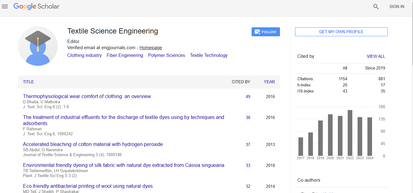
Journal of Textile Science & Engineering received 1008 citations as per Google Scholar report