Research Article - (2022) Volume 8, Issue 8
Received: 02-Aug-2022, Manuscript No. JPNP-22-71080;
Editor assigned: 05-Aug-2022, Pre QC No. JPNP-22-71080(PQ);
Reviewed: 22-Aug-2022, QC No. JPNP-22-71080;
Revised: 03-Oct-2022, Manuscript No. JPNP-22-71080(R);
Published:
11-Oct-2022
, DOI: 10.37421/2472-0992.2022.8.200
Citation: Moges, Getachew and Mengesha, Yohannes. "In Vivo
Antidiabetic Activity of Methanolic Extracts of Calpurnia Aurea and Bidens
Macroptera in Streptozotocin Induced Diabetic Mice." J Pharmacogn Nat
Prod8 (2022): 200.
Copyright: © 2022 Moges G, et al. This is an open-access article distributed under the terms of the creative commons attribution license which permits unrestricted
use, distribution and reproduction in any medium, provided the original author and source are credited.
Background: Diabetes mellitus is one of the most common chronic health problems globally. There is no satisfactory effective therapy to cure diabetes mellitus. Currently available drugs for managing diabetes produce some serious side effects and have decreased efficacy over time. Calpurnia aurea and Bidens macroptera have been used traditionally for the treatment of diabetes mellitus and other ailments in Ethiopia.
Objective: To investigate the antidiabetic activities of Bidens macroptera and Calpurnia aurea in streptozotocin induced diabetic mice.
Methods: Qualitative phytochemical screening tests were conducted to identify the chemical constituents of the plants. Healthy swiss albino mice of either sex (25 g-30 g) with no prior drug treatment were used for the present study. An acute toxicity study was carried out according to the 2008 organization for economic cooperation and development guideline 425. The effects of extracts of the plants on fasting blood glucose level and body weight of diabetic mice were evaluated using repeated dose antidiabetic activity test model. Fasting blood samples were collected from the control and test groups weekly to monitor blood glucose levels. Changes in body weight were also recorded weekly.
Results: Phytochemical screening of both plants indicated the presence of flavonoids, alkaloids, saponins, tannins, and phenolic compounds that might contribute to the antidiabetic activity. The medium Lethal Doses (LD50) of both extracts were higher than 2 g/kg body weight. The extracts also reduced the elevated blood glucose levels and improved the body weight loss of streptozotocin induced diabetic mice.
Conclusion: The methanolic extracts of Bidens macroptera and Calpurnia aurea revealed blood glucose lowering activity and improved body weight loss of diabetic mice over the 14 days treatment period.
Bidens macroptera • Calpurnia aurea • Antidiabetic activity • Diabetes mellitus
B. macroptera: Bidens Macroptera; BMEl: Bidens Macroptera Methanolic Leaf Extract; C. aurea: Calpurnia aurea; CAEl: Calpurnia Aurea Methanolic Leaf Extract; CAEr: Calpurnia Aurea Methanolic Root Extract.
Diabetes is a serious, chronic disease that occurs either when the pancreas does not produce enough insulin or when the body cannot effectively use the insulin it produces [1]. It is characterized by hyperglycemia, abnormal lipid, and protein metabolism along with specific long term complications affecting the retina, kidney, and nervous system [2]. The World Health Organization (WHO) has estimated that diabetes will be one of the world leading causes of death and disability in the next quarter century. Plants containing flavonoids, terpenoids, alkaloids, and phenolic compounds have been reported to have an antidiabetic activity [3-6]. In Ethiopia, Calpurnia aurea (C. aurea) is traditionally used for the treatment of diabetes mellitus, diarrhea, syphilis, athletes foot, amoebiasis, giardiasis, malaria, hypertension, rabies, and B. macroptera (B.macroptera) is used for the treatment of diabetes mellitus, brain cancer, as antioxidant, stomach ailments, and to remove pus from infected wounds [7-12]. The objective of the present study was to evaluate the possible antidiabetic properties of C. aurea and B. macroptera to establish the claimed biological activities of the plants.
Currently, there is no satisfactory effective therapy to cure diabetes mellitus. In addition to this, currently available agents for mitigating diabetes such as insulin and various oral antidiabetic agents such as sulfonylureas, biguanides, and glinides produce some serious side effects and have decreased efficacy over time [13-15]. Thus, an alternative therapy is required.
C. aurea and B. macroptera are commonly used in traditional medicine by the Agew Awi and Mecha people in ethiopia for the treatment of diabetes mellitus. The present study was intended to investigate the potential of these plants in reducing elevated blood glucose levels in Streptozotocin (STZ) induced diabetic mice to establish the claimed biological activities of these plants.
B. macroptera, locally known as adey abeba (Amharic) is known only from ethiopia. It belongs to the family Ateraceae. It grows on rocky mountain slopes and in grasslands, forest margins, and open places in ericaceous scrub; rarely, it is found along roadsides. It is more commonly found at higher altitudes.
C. aurea belongs to the family fabaceae and in amharic, it is known as digita. It is a small, multi-stemmed tree, 3 m-4 m tall. It is widely distributed in africa from cape province to eritrea and occurs in Southern India.
Collection and preparation of the plant material
The leaves, seeds, and roots of C. aurea (Ait.) benth. have been collected from chagni town and the leaves of B. macroptera have been collected from dessie town. The plants were then identified and authenticated at the department of botany, wollo university, dessie, ethiopia, where a voucher specimens GM 001 and GM 002 were deposited for C. aurea (Ait.) benth. and B. macroptera, respectively. The plants have been washed and separated by organ parts and then dried in the shade under room temperature. The dried plant parts have been powdered for maceration. The coarse powder was kept in polyethylene bags at room temperature until used for extraction [16].
Experimental animals
Healthy swiss albino mice of either sex (25 g-30 g) with no prior drug treatment were used for the present study. Mice were purchased from the Ethiopian Health and Nutrition Research Institute (EHNRI) and have been fed a standard commercial pellet diet and water with ad libitum. Before commencing the experiment, the mice were acclimatized to the experimental environment for 7 days under standard environmental conditions of temperature, relative humidity, and 12 hour light/dark cycle. The care and handling of the mice were according to the international guidelines for the use and maintenance of experimental animals.
Preparation of extracting solvent
The solvent system to be used for extraction was prepared by mixing up pure methanol and distilled water in 80:20 (v/v) ratios, respectively.
Preparation of plant extract
The dried and powdered plant parts have been exhaustively extracted by macerating with 80% methanol separately at room temperature for 72 hrs. with occasional manual stirring. The macerate was remacerated until the extracts gave no coloration. Then the extracts were filtered with Watchman filter paper No 1 and evaporated to dryness under reduced pressure by rotavapor at 50 rpm. The concentrated 80% methanolic extracts were collected in vials and placed in an oven at 30°C to evaporate the methanol completely. Finally, the dried extracts were placed in screw cup vials with the proper label and kept in a refrigerator at 4°C for use in the next phases of the experiment [17].
Phytochemical screening
Standard procedures were used to identify the chemical constituents (major secondary metabolites) of the plants. Qualitative tests which were based on the observation of color formation that occurs due to the reaction of secondary metabolites with different standard reagents were conducted.
Test for alkaloids: 2 grams of dried powder of the extracts of the plants were mixed with 10 ml of 1% hydrochloric acid. The mixture was boiled for 30 minutes in a water bath using a test tube. Then the mixture was allowed to cool and filtered using cotton plague on the test tube. To 5 ml of the filtrate, 5 drops of Mayer’s reagent (Mercuric chloride+Potassium iodide in water) was added and the formation of yellowish precipitate was inspected.
Test for flavonoids: To 2 ml of the plant extract, 3 drops of sodium hydroxide solution were added. The acute yellow color formation that changed to colorless by the addition of 3 drops of sulphuric acid was inspected.
Test for saponins: 1 ml solution of the plant extract was diluted with distilled water to 20 ml and shaken in a graduated cylinder for 15 minutes. This was inspected for the formation of a honeycomb that persists at least for half an hour.
Test for phenols: To 5 ml filtrate of the plant extract, 1 ml of 1% ferric chloride solution was added. Formation of green-blue color was inspected.
Test for tannins: To 2 ml of the plant extract, 5 drops of 2% lead acetate solution were added and the mixture was inspected for the development of yellow or orange precipitate.
Test for terpenoids: To 0.5 g of plant extract, 2 ml of chloroform and 5 ml of concentrated sulphuric acid were carefully added to form a layer and observed for the formation of the reddish-brown interface.
Test for anthraquinones: 1 gram of the powdered plant extract was placed in a dry test tube and 20 ml of chloroform was added.
This was heated in a steam bath for 5 min. The extract was filtered while hot and allowed to cool. To the filtrate was added an equal volume of 10% ammonia solution. This was shaken and the upper aqueous layer was observed for bright pink coloration as indicative of the presence of anthraquinones.
Preparation plant extract solution for administration
200 mg and 400 mg of solvent free dried leaf and root extracts of the two plants were dissolved separately in 10 mL of 1% tween 80 to prepare a stock solution of 20 mg/mL and 40 mg/mL daily, respectively to prepare dose levels of 200 mg/kg/day and 400 mg/kg/ day, b.w. Then, the desired doses were administered according to the body weight of the mice in the respective groups. The doses were selected based on previous studies [18].
Acute toxicity study
Acute oral toxicity study was performed according to the 2008 Organization for economic cooperation and development 35 guideline 425. Five healthy female swiss albino mice aged 8 weeks-12 weeks selected by random sampling were used for this study. Female mice were used for the acute toxicity study because females are generally slightly more sensitive to toxicity. Before administration of extracts of the plants, the mice were fasted for 4 hrs. But provided only with water. Following the period of fasting, they were weighed and the plant extracts have been administered orally. The dose for each mouse was calculated according to the fasting body weight. First, a sighting study at a starting dose of 2000 mg/kg b.w was conducted on a single mouse to determine the starting dose. Since the mouse survived for 24 hrs. after the sighting study, an additional four mice were administered with the same dose of extracts of the plants. Then they were housed separately and observed continuously for the first 4 hrs. With a 30 min interval and then for 14 consecutive days with an interval of 24 hrs. for any visible toxic manifestations such as feeding behavior, hair erection, tremors (hyperactivity), lacrimation, diarrhea, sleep(sedation), coma, mortality, and hypothermia. All observations were systematically recorded, with individual records being maintained for each mouse [19].
Oral glucose tolerance test
For the oral glucose tolerance test and the repeated antidiabetic activity test models, the leaf extract of B. macroptera and root extract of B. macroptera were used.
Normal nondiabetic mice, which fasted for 6 hrs. were randomly divided into six experimental groups (n=6).
• Group I: Treated with 10 ml/kg b.w of 1% Tween 80 (Negative control).
• Group II: Treated with 10 mg/kg b.w of glibenclamide (Positive control).
• Groups III and IV: Treated with 200 mg/kg b.w of each of C. aurea (Ait.) benth. and B. macroptera, separately.
• Groups V and VI: Treated with 400 mg/kg b.w of each of C. aurea (Ait.) benth. and B. macroptera, separately.
Fasting blood samples were taken from the mouse tail vein of all groups and blood glucose levels were measured just before the administration of the plant extract, the standard drug, and the vehicle.
After 30 minutes of extract, standard drug, and vehicle administration, the mice of all groups were orally treated with 2 g/kg b.w of glucose.
Then Blood samples were collected again from all groups at 30 min, 60 min and 120 min of intervals after glucose loading, and blood glucose levels were measured.
Preparation of 0.1 molar citrate buffer
Citric acid (10.5 g) and sodium citrate (14.7 g) were accurately weighed and mixed in 50 ml of distilled water. The volume was made up to 1000 ml with distilled water and the pH was adjusted to 4.5 by sodium hydroxide solution.
Induction of diabetes in experimental animals
Mice were made diabetic by administering a single intraperitoneal (i.p) injection of a freshly prepared solution of STZ to overnight fasted mice. 20 mg/ml stock solution of STZ was prepared just before administration (stability) using 0.1 M citrate buffer, pH 4.5. Then a single i.p injection of 200 mg/kg b.w of STZ was administered to each mouse. The negative control mice were injected with the same concentration of citrate buffer only (vehicle). Since STZ is capable of inducing fatal hypoglycemia because of massive pancreatic insulin release, the mice were given food ad libitum and 5% glucose solution in a feeding bottle 1 hr after STZ administration overnight to overcome the initial drug induced hypoglycemic (early hypoglycemic phase) mortality. Then the mice were allowed to stabilize for 3 days after STZ injection and screened for diabetes by taking blood samples from the tail vein on the 3rd day. Those mice having a fasting blood glucose level above 200 mg/dL were considered as diabetic and were eligible for inclusion in the study. The mechanism by which STZ induces diabetes mellitus includes selective destruction of insulin-secreting pancreatic β-cells and causes poor glucose uptake by peripheral tissues. Treatment with extracts of the plants and standard drug was started on the 3rd day after STZ injection and this day was considered as day 0th of treatment. Then all mice were given water and food ad libitum [20].
Anti-diabetic activity test
Diabetic and nondiabetic mice were randomized into seven groups (n=6) and received the following treatment.
• Group I: Non-diabetic mice, received 10 ml/Kg b.w of 1% tween 80 (normal control)
• Group II: Diabetic mice received 10 ml/Kg b.w of 1% tween 80 (diabetic control)
• Groups III and IV: Diabetic mice received 200 mg/kg b.w of extracts of the plants separately.
• Groups V and VI: Diabetic mice received 400 mg/kg b.w of extracts of the plants separately.
• Group VII: Diabetic mice received 10 mg/kg b.w of glibenclamide, standard drug.
The extracts of the plants, the standard drug, and the vehicle were administered daily for 14 consecutive days in the morning by gastric intubation with oral gavage. Stock solutions of 20 mg/ml and 40 mg/ml of extracts of the plants and 1 mg/ml of glibenclamide in 1% Tween 80 were prepared freshly before administration each day.
Measuring fasting blood glucose level
Fasting blood samples were collected weekly on the 0th, 7th, and 14th days of treatment from the tail vein of the overnight fasted mice, 1 hr after administration of the extracts, the standard drug, and the vehicle to monitor blood glucose levels. Blood glucose level was measured using a blood glucose meter.
Determination of weight of experimental mice
Bodyweight gain or loss in each experimental mouse was measured and recorded weekly on the 0th, 7th, and 14th days of treatment.
Data analysis
Data were analyzed using SPSS version 26.0. All values of fasting blood glucose level and body weight were presented as mean ± standard deviation. Mean differences between controls and the different test groups were compared for statistical significance using one-way ANOVA followed by the post-hoc dunnett’s multiple comparisons. Mean values were considered significantly different if P value <0.05 or as very significantly different if P value <0.01.
Percentage yields of the plants
The percentage yields (% w/w) of different parts of the plants have been determined. The leaves of B. macroptera gave a higher percentage yield than the stems. Similarly, the leaves of C. aurea gave a higher percentage yield than its seeds (Table 1).
| Plant part | Dried weight of plant part (gm) | Weight of crude extract (gm) | % Yield |
|---|---|---|---|
| Leaves of B.macroptera | 60 | 6.3 | 10.5 |
| Stems of B.macroptera | 60 | 4.2 | 7 |
| Leaves of C.aurea | 65 | 7.15 | 11 |
| Seeds of C.aurea | 50 | 4.7 | 9.4 |
| Roots of C.aurea | 67 | 6.6 | 9.85 |
Phytochemical constituents
The phytochemical investigation of the leaf extracts of B. macroptera revealed the presence of alkaloids, flavonoids, saponins, and terpenoids and the absence of phenols, tannins, and anthraquinones. And, the qualitative phytochemical analysis of the leaves, seed, and root extracts of C. aurea showed that alkaloids, flavonoids, saponins, phenols, tannins, and terpenoids are present in the leaves, roots and seeds of C. aurea whereas, anthraquinones are absent (Table 2).
| Constituents | Test procedure | Indicator | Test result | |
|---|---|---|---|---|
| B. macroptera | C. aurea | |||
| Alkaloids | Filtrate+Mayer's reagent | Yellowish precipitate | +++ | +++ |
| Flavonoids | 2 ml extract+3 drops of NaOH | Yellow color that clears on adding HCl | +++ | +++ |
| Phenols | 5 ml extract+1% FeCl3+1 mL of K3Fe (CN)6 | Green blue color | _ _ | +++ |
| Saponins | 1 ml extract+distilled water+shake | Formation of foam | +++ | +++ |
| Tannins | 2 ml extract+5 drops of 2% lead acetate | Yellow or orange precipitate | _ _ | +++ |
| Terpenoids | 0.5 g extract+2 ml chloroform+5 ml H2SO4 | Reddish brown interface | +++ | +++ |
| Anthraquinones | 1 g powder+20 ml chloroform +10% ammonia | Bright pink layer | _ _ | _ _ |
Acute toxicity study
As shown in Table 3, the absence of mortality in the test groups over the 14 days following oral administration of the extracts at 2 g/kg b.w dose indicates the median Lethal Dose (LD50) of the extracts of the plants is higher than 2 g/kg b.w. According to OECD guidelines for acute oral toxicity, an LD50 dose of 2 g/kg and above is categorized as unclassified and hence extracts of the plants are safe.
Since extracts of the plants were safe at a dose of 2 g/kg, 400 mg/kg was chosen as the maximum dose for the antidiabetic model.
| Mouse ID | Outcome | Signs of toxicity | ||
|---|---|---|---|---|
| B.macroptera | C.aurea | B.macroptera | C.aurea | |
| 1 | Survived | Survived | None | None |
| 2 | Survived | Survived | None | None |
| 3 | Survived | Survived | None | None |
| 4 | Survived | Survived | None | None |
| 5 | Survived | Survived | None | None |
Oral glucose tolerance test
Administration of 2 g/kg b.w glucose significantly (p<0.01) raised the blood glucose level of all groups after 30 min. The elevated blood glucose level of the negative control mice remained high over the next 90 min. The blood glucose levels of the groups treated with leaf extract of B. macroptera and root extract of C. aurea reduced to near normal or basal values after 1 hr, while glibenclamide 10 mg/kg brought the elevated blood glucose level to near normal or basal value after 30 min (p<0.01). 120 min after oral glucose loading, there were no statistically significant differences in blood glucose levels when the glibenclamide treated group was compared with the groups treated with leaf extract of B. macroptera and root extract of C. aurea and when the plant extract treated groups were compared with each other, whereas significant differences were observed when the negative control was compared with glibenclamide treated group and with the plant extract treated groups (Figure 1).

Figure 1. Antihyperglycemic effect of extracts of B. macroptera and C. aurea in glucose loaded hyperglycemic mice.
NEC-Negative control, Glb-Glibenclamide 10 mg/kg, BMEl 200-B.
Macroptera leaf extract 200 mg/kg, BMEl 400-B. Macroptera leaf extract 400 mg/kg, CMEr 200-C.aurea root extract 200 mg/kg, CMEr 400-C. aurea root extract 400 mg/kg.
Anti-diabetic activity test
On day 0th of treatment, the fasting blood glucose levels of all diabetic mice were significantly (P<0.01) higher than those of normal control mice. All diabetic groups receiving extracts of the two plants showed a statistically significant (P<0.01) reduction in fasting blood glucose levels on days 7th and 14th of treatment compared with their respective baseline blood glucose levels. There was no significant (P>0.05) difference in fasting blood glucose levels of normal control mice over the 14 days of treatment, whereas the diabetic control mice showed a significant (P<0.01) increase in fasting blood glucose level at day 14th. There was a statistically significant (P<0.01) difference between each dose of B. macroptera extract on days 7th and 14th of treatment. However, there were no significant (P>0.05) differences between each dose of C. aurea. Except the group receiving 400 mg/kg b.w of B. macroptera extract, there was no significant (P>0.05) difference between diabetic mice receiving extracts of the plants.
The 200 mg/kg b.w dose of B. macroptera extract produced a reduction of 21.39% and 22.93% on fasting blood glucose levels of diabetic mice on days 7th and 14th, respectively while, the 400 mg/ kg b.w dose reduced the fasting blood glucose level by 33.25% and 33.80% on days 7th and 14th, respectively. Treatment with 200 mg/kg b.w of extract of C. aurea resulted in 19.89% and 21.48% reduction in fasting blood glucose level of diabetic mice on days 7th and 14th, respectively, whereas 400 mg/kg b.w dose resulted in 27.72% and 30.42% reduction of fasting blood glucose level on days 7th and 14th, respectively. Glibenclamide treated diabetic mice showed 56.84%and 57.73% reduction on days 7th and 14th, respectively, as a positive control (Table 4).
| Group | Dose (mg/kg) | Fasting blood glucose level (mg/dl) | Reduction in fasting blood glucose | |||
|---|---|---|---|---|---|---|
| Baseline | 7th day | 14th day | 7th day | 14th day | ||
| Normal control | Vehicle | 72.47 ± 2.91b | 74.35 ± 4.24b | 74.67 ± 5.48 a b | -2.59% | -3.04% |
| Diabetic control | Vehicle | 381.80 ± 1.70a | 384.92 ± 2.49a | 389.85 ± 2.86aγ | -0.82% | -2.11 |
| Glibenclamide | 10 | 378.47 ± 3.19a | 163.33 ± 8.87abγ | 159.99 ± 8.09abγ | 56.84% | 57.73% |
| Diabetic+BME | 200 | 344.23 ± 2.92ab | 270.58 ± 4.49abγ | 265.23 ± 4.40abγ | 21.39% | 22.93% |
| Diabetic+BME | 400 | 367.75 ± 2.13ab | 245.48 ± 3.37abγ | 243.45 ± 4.22abγ | 33.25% | 33.80% |
| Diabetic+BME | 200 | 340.03 ± 3.68ab | 272.39 ± 3.02abγ | 266.99 ± 4.79abγ | 19.89% | 21.48% |
| Diabetic+CME | 400 | 381.58 ± 6.59a | 275.82 ± 4.49abγ | 265.50 ± 5.61abγ | 27.72% | 30.42% |
Each value represents the mean ± SD (n=6). a p<0.01 compared with normal control values,ᵦ p<0.01 compared with diabetic control values and γ p<0.01 compared with baseline values (day 0) of fasting blood glucose level of the mice of each group. BMEl- B. macroptera leaf extract, CMEr – C. aurea root extract.
Effect of B. macroptera and C. aurea extracts on weight reduction in STZ induced diabetic mice: Bodyweight gain or loss in each mouse was measured and recorded weekly on days 0th, 7th, and 14th of treatment. There were no significant (P>0.05.) differences in the body weights of normal control mice on days 7th and 14th compared to their initial body weights. However, the body weights of untreated diabetic control mice were significantly reduced on days 7th and 14th compared to their initial weights (P<0.01). Diabetic mice treated with the crude extracts did not show significant (P>0.05) differences in body weight at all doses compared to their baseline body weight on day 14th which is comparable with the standard drug. The weight gain of diabetic mice treated with 200 mg/Kg BME, 400 mg/Kg BME, 200 mg/kg CAE, 400 mg/kg CAE and the standard drug were 0.46%, 2.62%, 0.6%, 1.23% and 5%, respectively on day 14th (Figure 2).

Figure 2. Effect of extracts of B. macroptera and C. aurea on body weight of diabetic mice.
NC-Normal Control, DC-Diabetic Control, Glb-Glibenclamide 10 mg/kg, BMEl 200-B. macroptera leaf extract 200 mg/kg, BMEl 400-B. macroptera leaf extract 400 mg/kg, CMEr 200 -C. aurea root extract 200 mg/kg, CMEr 400-C.aurea root extract 400 mg/kg.
The present study was designed to investigate the antidiabetic activity of hydroalcoholic extracts of B. macroptera and C. aurea in STZ induced diabetic mice. Diabetes mellitus was induced in the mice by intraperitoneal administration of 200 mg/Kg b.w STZ. The mechanism by which STZ causes diabetes mellitus includes selective destruction of insulin secreting pancreatic β-cells and causes poor glucose uptake by peripheral tissues. Both doses of extracts of the plants showed a significant antihyperglycemic effect during the 14 days of treatment in a dose dependent manner.
The mechanism of action of the crude extracts of the two plants might be due to an insulin mimetic effect on muscle and adipose tissue by either stimulating glucose uptake and metabolism, by inhibiting hepatic gluconeogenesis and glycogenolysis, by stimulation of regeneration process or increase pancreatic secretion of insulin from existing β-cells and/ or inhibition activity against α-glucosidase enzymes in the small intestine which convert disaccharides into monosaccharides for sake of absorption.
The blood glucose lowering effect of these crude extracts may be due to the presence of different antioxidant phytochemicals such as flavonoids, alkaloids, polyphenols, terpenoids, and tannins, which act as free radical scavengers. Flavonoids are known to have insulinogenic and pancreatic beta-cell regenerating activities.
Among the extracts of the plants, a maximal reduction in blood glucose level was observed at a dose of 400 mg/kg b.w of B. macroptera extract. This might be attributed to the 400 mg/kg dose of B. macroptera extract contained a higher amount of active component (s) responsible for antidiabetic activity.
Groups receiving the vehicle showed slight increments in blood glucose levels during the 14 days of treatment compared to their baseline values. However, significant (P<0.01) reductions in fasting blood glucose levels were observed in both the OGTT and STZ induced diabetic models after the administration of the extracts of both plants and the standard drug. This indicates reductions in blood glucose levels are attributed to treatment.
The present study also revealed that STZ caused significant body weight loss in diabetic control mice. The loss of body weight in untreated diabetic mice is due to increased muscle wasting 58 and catabolism of tissue proteins. Hydroalcoholic extracts of B. macroptera and C. aurea prevented weight loss. Weight loss is characteristic of diabetic patients. The weight gain of groups treated with extracts of the plants and the standard drug in STZ induced diabetic mice evidences the antidiabetic activity of these medicinal plants.
The acute toxicity study indicated that the crude extracts of both plants are nontoxic under observable conditions in mice. The crude extracts of B. macroptera and C. aurea possessed most of the bioactive constituents used for the management of diabetes mellitus. Extracts of the plants enhanced glucose tolerance in the mice and had substantial antidiabetic effects at doses of 200 and 400 mg/kg b.w. The crude plant extracts also protected massive body weight loss. Therefore, the phytochemical constituents of the plants might be useful in the prevention of diabetic complications and may play a significant role as an alternative for the management of diabetes mellitus.
Further pharmacological and biochemical investigations have to be done to explore the exact mechanism of action of the extracts.
Further quantitative phytochemical analysis and fractionation should be done to isolate and identify the active component (s) responsible for the antidiabetic activity of the plants. Other parts of the plants should be screened for antidiabetic activity. Blood and serum biochemical parameters such as glycated hemoglobin, insulin level, and histopathology of pancreas, liver, and kidney organs have to be done.
The authors are grateful to Wollo University for its financial support.
The study was supported by a research grant from wollo university; dessie, ethiopia. The funder had no role in the design of the study and collection, analysis, and interpretation of data and in writing the manuscript.
All datasets used and analyzed during the present study are available from the corresponding author on reasonable request.
Ethical review board of college of medicine and health sciences has approved the protocol. The care and handling of the mice were according to the international guidelines for the use and maintenance of experimental animals.
Both authors contributed to the data analysis, drafting, and revising the article, and gave final approval of the version to be published. GM conducted the actual experiment and statistical analysis. YM reviewed the proposal and involved in all implementation stages of the study and write up. Both authors reviewed and approved the final version of the manuscript.
The authors declare that they have no competing interests.
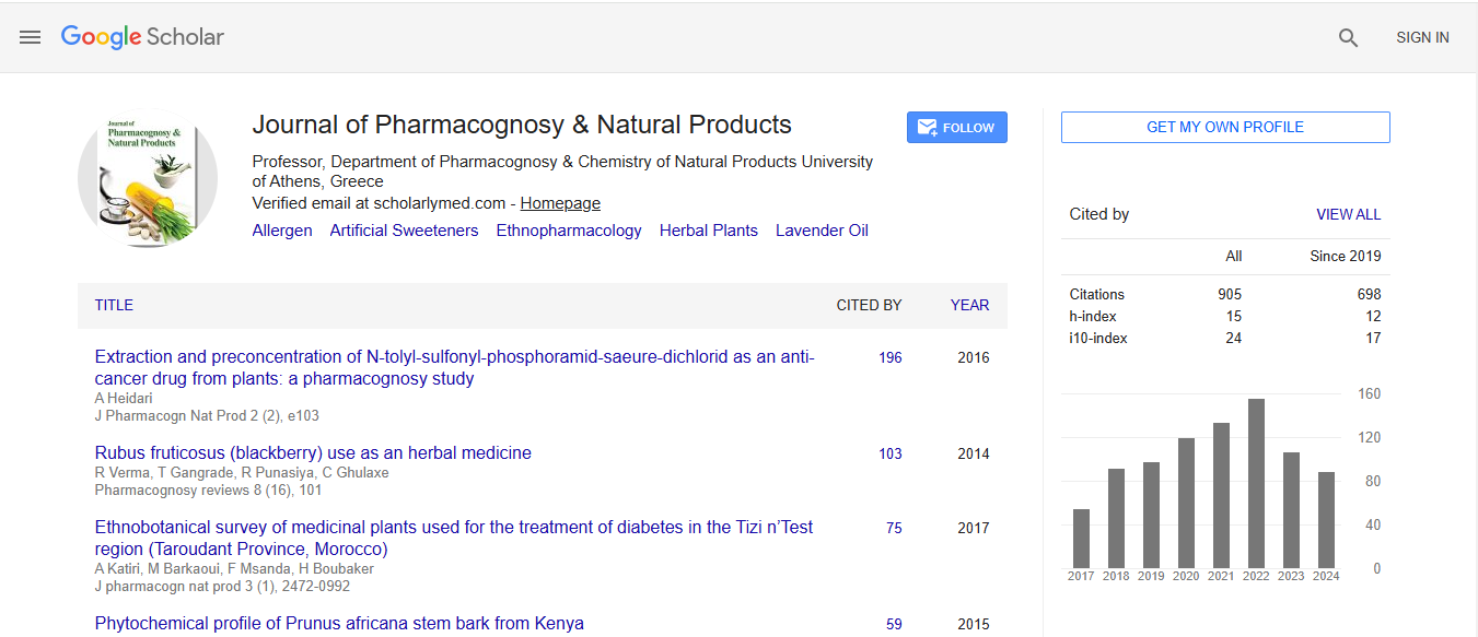
Journal of Pharmacognosy & Natural Products received 606 citations as per Google Scholar report