Short Communication - (2025) Volume 10, Issue 3
Received: 01-May-2025, Manuscript No. JPNM-26-185774;
Editor assigned: 05-May-2025, Pre QC No. P-185774;
Reviewed: 19-May-2025, QC No. Q-185774;
Revised: 22-May-2025, Manuscript No. R-185774;
Published:
29-May-2025
, DOI: 10.37421/2472-100X.2025.10.349
Citation: Mehta, Arjun. ”Advanced Neuroimaging Revolutionizes Pediatric Brain Disorder Care.” J Pediatr Neurol Med 10 (2025):349.
Copyright: © 2025 Mehta A. This is an open-access article distributed under the terms of the Creative Commons Attribution License, which permits unrestricted use, distribution and reproduction in any medium, provided the original author and source are credited.
Neuroimaging techniques have fundamentally transformed the landscape of pediatric brain disorder diagnosis and management. These advanced modalities, including MRI, fMRI, DTI, and MRS, provide unparalleled insights into the intricate structure, dynamic function, and complex connectivity of the developing brain. This detailed understanding is crucial for the early identification, precise localization of lesions, and the tailoring of personalized treatment strategies for a wide spectrum of conditions. The ability to gain such in-depth information is paramount for conditions as varied as developmental abnormalities, epilepsy, brain tumors, and traumatic brain injuries, ultimately contributing to improved patient outcomes and more accurate prognoses. [1] Diffusion tensor imaging (DTI) has emerged as a pivotal tool in assessing the integrity of white matter tracts within the pediatric brain. It possesses the remarkable capability to detect subtle microstructural alterations that may not yet manifest as overt clinical symptoms. Furthermore, DTI is instrumental in tracking the efficacy of ongoing treatments and elucidating the long-term neurological consequences of brain injury in children. [2] Functional MRI (fMRI) offers critical insights into the activity of brain networks and the functional organization of the pediatric brain. This technique is particularly valuable in characterizing neurodevelopmental disorders such as autism spectrum disorder and ADHD. Comprehending the intricate ways in which different brain regions communicate and interact is essential for the development of effective, targeted interventions. [3] Magnetic Resonance Spectroscopy (MRS) enables the non-invasive quantification of key brain metabolites. This capability is invaluable for the diagnosis and ongoing monitoring of metabolic disorders. It also aids in understanding the biochemical shifts that accompany various pediatric brain diseases, providing a window into the cellular health of the brain. [4] The integration of sophisticated neuroimaging with genetic and clinical data is progressively gaining importance for achieving a comprehensive understanding of complex pediatric brain disorders. This synergistic, multimodal approach facilitates more accurate diagnoses and paves the way for the development of precision medicine strategies tailored to individual patients. [5] Neuroimaging techniques play an indispensable role in the early detection and diligent monitoring of brain tumors in children. Advanced MRI sequences, particularly those employing contrast enhancement and diffusion-weighted imaging, are vital for accurately characterizing tumors, planning surgical interventions, and assessing the effectiveness of treatment regimens, thereby significantly impacting survival rates. [6] In the management of pediatric epilepsy, neuroimaging is indispensable for identifying the underlying structural causes of seizures, guiding surgical decision-making, and monitoring seizure activity. Cutting-edge MRI sequences and PET scans contribute significantly to a more precise localization of seizure foci, enabling more effective treatment. [7] The management of traumatic brain injury (TBI) in children is substantially enhanced by serial neuroimaging assessments. These evaluations help in tracking the evolution of initial lesions, detecting secondary injuries such as edema or hemorrhage, and monitoring the impact of therapeutic interventions. Advanced imaging methods can reveal subtle damage that might be missed on initial scans. [8] Quantitative neuroimaging techniques, including volumetric analysis and tractography, are increasingly providing objective measures of brain development and the specific impact of various disorders on brain structure and connectivity in pediatric populations. These quantitative insights are proving vital for both research endeavors and daily clinical practice. [9] The application of artificial intelligence (AI) and machine learning (ML) to pediatric neuroimaging data holds immense potential for refining diagnostic accuracy, predicting treatment responses, and identifying novel biomarkers for the early detection of brain disorders. This technological advancement is poised to revolutionize the approach to managing these complex conditions. [10]
Neuroimaging techniques have revolutionized the diagnosis and management of pediatric brain disorders, offering unprecedented insights into brain structure, function, and connectivity. Advanced modalities like MRI, fMRI, DTI, and MRS are crucial for early detection, precise localization of lesions, and personalized treatment strategies across a spectrum of conditions, from developmental abnormalities and epilepsy to tumors and traumatic brain injuries, ultimately improving patient outcomes and informing prognosis. [1] Diffusion tensor imaging (DTI) is instrumental in evaluating white matter integrity in pediatric neurological conditions. It effectively reveals subtle microstructural changes that may precede overt clinical symptoms, and its application extends to tracking the effects of treatment and understanding the long-term consequences of brain injury in children. [2] Functional MRI (fMRI) provides critical insights into brain network activity and functional organization in children, aiding in the characterization of disorders like autism spectrum disorder and ADHD. Understanding how brain regions communicate and interact is essential for developing targeted interventions. [3] Magnetic Resonance Spectroscopy (MRS) allows for the non-invasive quantification of brain metabolites. This capability is invaluable in diagnosing and monitoring metabolic disorders and understanding the biochemical changes associated with various pediatric brain diseases, offering a window into the cellular health of the brain. [4] The integration of advanced neuroimaging with genetic and clinical data is becoming increasingly important for a comprehensive understanding of complex pediatric brain disorders. This multimodal approach facilitates more accurate diagnoses and the development of precision medicine strategies. [5] Neuroimaging plays a vital role in the early detection and monitoring of brain tumors in children. Techniques like MRI with contrast enhancement and diffusion-weighted imaging help in tumor characterization, surgical planning, and assessment of treatment response, significantly impacting survival rates. [6] In the context of pediatric epilepsy, neuroimaging is crucial for identifying underlying structural abnormalities, guiding surgical interventions, and monitoring seizure activity. Advanced MRI sequences and PET scans contribute to a more precise understanding of seizure foci. [7] The management of traumatic brain injury (TBI) in children benefits significantly from serial neuroimaging, which helps track the evolution of lesions, assess for secondary injuries like edema or hemorrhage, and monitor the effects of interventions. Advanced techniques can reveal subtle damage not apparent on initial scans. [8] Quantitative neuroimaging techniques, such as volumetric analysis and tractography, are providing objective measures of brain development and the impact of various disorders on brain structure and connectivity in children. These quantitative insights are vital for research and clinical practice. [9] The application of artificial intelligence and machine learning to neuroimaging data in pediatrics holds immense promise for improving diagnostic accuracy, predicting treatment response, and identifying novel biomarkers for early detection of brain disorders. This is set to transform how we approach these conditions. [10]
Neuroimaging techniques such as MRI, fMRI, DTI, and MRS are revolutionizing the diagnosis and management of pediatric brain disorders. These advanced methods provide detailed insights into brain structure, function, and connectivity, enabling early detection, precise localization of lesions, and personalized treatment plans for conditions like developmental abnormalities, epilepsy, tumors, and traumatic brain injuries. DTI specifically evaluates white matter integrity, while fMRI maps brain network activity. MRS quantifies brain metabolites, aiding in metabolic disorder diagnosis. Integrating neuroimaging with genetic and clinical data supports precision medicine. Techniques are also vital for brain tumor characterization, surgical planning, and assessing treatment response. In epilepsy, neuroimaging helps identify seizure foci and guides interventions. For TBI, serial imaging tracks lesion evolution and secondary injuries. Quantitative analyses offer objective measures of brain development and disorder impact. The future of pediatric neuroimaging is bright with the integration of AI and machine learning to enhance diagnostic accuracy and predictive capabilities.
None
None
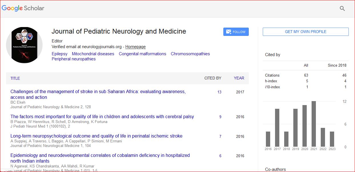
Journal of Pediatric Neurology and Medicine received 68 citations as per Google Scholar report