DOI: 10.4172/2167-0390.1000e118
DOI: 10.4172/2167-0390.1000e119
Harris M (2013) Nutrient Interaction: Are Lutein and Omega-3 Docosahexaenoic Acid (DHA) Conditionally Essential and Complementary Nutrients for Visual Function? Vitam Trace Elem 2:e119.
DOI: 10.4172/2167-0390.1000e120
DOI: 10.4172/2167-0390.1000e121
Carly J Moss and Suresh T Mathews
DOI: 10.4172/vms.1000111
The water soluble B vitamin, thiamin, in its coenzyme form as thiamin pyrophosphate (TPP), is necessary for key reactions in glucose metabolism. For this reason, associations between thiamin status or thiamin supplementation and diabetes have been the focus of recent research. Currently, it is not clear how occurrence of diabetes relates to parameters of thiamin status, such as plasma thiamin levels. However, there is strong evidence that the diabetic state increases urinary thiamin excretion and decreases the activity of transketolase (TK), a TPP-dependent enzyme of the hexose monophosphate (HMP) shunt in various body tissues. Impairment of TK activity and subsequent downregulation of the HMP shunt activates several pathways that contribute to vascular damage and development of diabetes-related comorbidities such as retinopathy, cardiomyopathy, and nephropathy. Thiamin supplementation has been shown to be effective in restoring TK activity in animal models of diabetes, and in type 1 and type 2 diabetic individuals. Thiamin supplementation has also been shown to be effective in preventing or reversing, either partially or completely, hyperglycemia-induced damage to vascular endothelial cells, and microalbuminuria associated with diabetic nephropathy. Here, we review from current literature examining the relationship between diabetes and thiamin status as well as intervention trials evaluating the effect of thiamin supplementation on glycemic control and prevention of vascular comorbidities of diabetes.
James C Barton, Paul C Adams, Ronald T Acton, Mark Speechley, Christine E McLaren, Gordon D McLaren, Victor R Gordeuk and John H Eckfeldt
DOI: 10.4172/vms.1000112
In non-screening hemochromatosis patients with HFE C282Y homozygosity who achieve iron depletion, the proton pump inhibitor (PPI) omeprazole decreases non-heme iron absorption from test meals and decreases maintenance phlebotomy requirements. We sought to determine the effect of taking PPIs and histamine H2 receptor antagonists (H2RAs) on serum ferritin (SF) levels in C282Y homozygotes diagnosed in a screening study. Methods: We compared mean ln SF in 171 homozygotes (60 men, 111 women) who reported taking and not taking PPIs and H2RAs. We performed linear regression on ln post-screening SF using age, sex, reports of taking PPIs and H2RAs, and free thyroxine levels. Results: Eleven homozygotes (6.4%) took PPIs; twelve (6.7%) took H2RAs. Mean ln SF values of male and female homozygotes who took PPIs were ~one-half those of homozygotes without PPI reports, but the differences were not significant. In an initial five-factor regression model, H2RAs and free thyroxine levels were not significant predictors of ln SF. In a final three-factor regression model, taking PPIs was associated with lower ln SF (p=0.0031) after controlling for age and sex. The final model explained 31% of the variance in ln SF. Use of H2RAs was not independently associated with ln SF. Conclusions: Taking PPIs is associated with lower SF levels in HFE C282Y homozygotes diagnosed in screening. Inorganic iron absorption may be decreased in some homozygotes who take PPIs; others who take PPIs may have upper gastrointestinal lesions that lower SF due to blood loss.
Isabelle Germain, Sherry Agellon and Hope Weiler
DOI: 10.4172/vms.1000113
Vitamin D is important to bone health. This study examined vitamin D intake and status in institutionalized elderly men in relation to biomarkers of bone metabolism and functional indicators. Materials and Methods: Elderly male veterans were studied in Phase I (n=40) for 16 weeks (April, June, August 2008) and Phase II (n=30) for another 16 weeks (October and December 2008 and February 2009) for dietary vitamin D using 5 day menu selection (Phase I) and using 3×3-d weighed food records (Phase II). Anthropometric data, Mini-Mental State Evaluation (MMSE) scores and sun exposure were collected. Functional capacity was assessed using the Frail Elderly Functional Assessment Tool (FEFA) and handgrip strength. Biochemistry included serum 25-hydroxyvitamin D (25(OH)D), parathyroid hormone (PTH), osteocalcin (OC) and C-terminal telopeptides of Type 1 collagen (CTX). Mixed model ANOVA and Pearson correlations analyses were used. Results: Participants were relatively healthy (Age: 85 ± 3 years (Mean ± SD), BMI: 26.1 ± 4.3 kg/m2, MMSE: 25 ± 5, FEFA: 13 ± 8, grip strength: 22 ± 8 kg). Sixty-six percent (280 ± 120 IU) of the planned dietary vitamin D was consumed. Vitamin D came mainly from fortified milk and meal supplements and 33% took pill supplements (400-800 IU/d). Serum 25(OH) D concentration rose by summer (Phase I: 60.9 ± 24.4, 68.2 ± 24.6 and 76.1 ± 22.4 nmol/L, respectively) and declined thereafter (Phase II: 57.7 ± 24.1, 62.9 ± 30.7 and 61.3 ± 29.2 nmol/L). PTH was lower in spring compared to late summer through winter whereas CTX and OC did not change. Serum 25(OH) D was correlated to BMI, but not to indicators of functional status. Conclusions: In long-term care, vitamin D from foods and supplements fails to meet recommendations of 800 IU (20 μg) for those over 70 y.
Nittaya Chansiw, Kanjana Pangjit, Wirote Tuntiwechapikul, Chada Phisalaphong, Suthat Fucharoen, John B. Porter and Somdet Srichairatanakool
DOI: 10.4172/vms.1000114
An interruption of the iron metabolism with chelators can lead to a significant inhibition of cancer cell growth. 1-(N-acetyl-6-aminohexyl)-3-hydroxy-2-methylpyridin-4-one or CM1, is a novel synthetic bidentate iron chelator which was successfully synthesized by our group. We have studied the characteristics and iron-chelating activity of this compound. Nevertheless, the anti-cancer activity of the chelator is largely unknown. In this study, we demonstrated the cytotoxicity and apoptogenic activity of CM1 against human leukemic cell lines-HL-60 and U937. 3-(4,5-Dimethylthiazol- 2-yl)-2,5-diphenyltetrazolium bromide (MTT) assay was performed for the cytotoxicity study. The results showed that CM1 inhibited the cell growth and metabolic activity of the leukemic cells. Flow cytometric analysis clearly demonstrated the dose and time-response of CM1-induced apoptosis in these two cells. CM1 arrested the cell populations in the sub G1 phase after 24 hours of exposure. The cancer cells induced by the compound significantly decreased mitochondria membrane potential (Δψm), and increased the activation of caspase-2,-3,-8 and caspase-9 activities. Possibly, CM1 would interact with nonheme iron-containing enzymes, such as ribonucleotide reductase and depleting intracellular iron essential for fast dividing cancer cells, leading to cell apoptosis. The CM1 may act as a reducing agent and help to maintain the CM1-Fe2+ complex which can generate radicals.
Kanokwan Kulprachakarn, Kanjana Pangjit, Chada Phisalaphong, Suthat Fucharoen, Robert C. Hider and Somdet Srichairatanakool
DOI: 10.4172/vms.1000115
Iron overload associated with oxidative stress is a serious problem in transfusion-dependent patients with β-thalassemia major. The increased iron overload in several organs may be caused by higher intestinal absorption along with less intensive chelation therapy. Liver iron overload could in turn facilitate the development or persistence of chronic progressive liver disease. Previous studies have shown that chelation with desferrioxamine (DFO) and deferiprone (DFP) substantially reduced body-iron scores in β-thalassemia patients with transfusional iron overload. We have synthesized and characterized a new bidentate iron chelator, 1-(N-acetyl-6-aminohexyl)-3-hydroxy-2- methylpyridin-4-one (CM1). The compound can efficiently scavenge iron from both ferrous and ferric salts and plasma non-transferrin bound iron (NTBI). In this study we have studied the efficacy of the CM1 treatment on the decrease of levels of the labile iron pool (LIP) and reactive oxygen species (ROS) in iron-loaded mouse hepatocyte and HepG2 cell cultures. The isolated hepatocytes were treated with DFP, DFO and CM1 at different concentrations. The treated cells were analyzed for intracellular LIP using the calcein fluorescent technique and ROS levels using the dichlorofluorescein (DCF) fluorescent method. It was found that CM1 reduced the levels of intracellular LIP and hydrogen peroxideinduced ROS in both treated cells in a concentration-dependent manner. The combination treatment of CM1 with 25 μM DFP and DFO was demonstrated to decrease the levels of the LIP in both cells and tended to reduce the levels of ROS in HepG2 cells. Our findings support the evidence of iron-chelating and free radical-scavenging activities of CM1 in the livers with iron overload, which potentially can protect against oxidative liver inflammation and fibrosis. The efficacy of the CM1 treatment needs to be further investigated intensively under in vivo conditions.
Nittaya Chansiw, Kanjana Pangjit, Chada Phisalaphong, Suthat Fucharoen, Patricia Evans, John B Porter and Somdet Srichairatanakool
DOI: 10.4172/vms.1000116
Deferiprone (DFP) (MW=139 Da, Kpart=0.11) is an effective iron chelator used for the treatment of iron overload in thalassemia patients, but the drug is not free from side effects. We have synthesized a novel oral bidentate iron chelator, 1-(N-acetyl-6-aminohexyl)-3-hydroxypyridin-4-one (CM1) (MW=256 Da, Kpart=0.53), which is an analogue of DFP. This compound is more lipophilic than DFP and can bind iron efficiently. Our current results have demonstrated that CM1 reduced iron-induced redox damage and decreased levels of the intracellular iron pool (LIP) in cultured hepatocytes, effectively. However, the toxicity of CM1 remains largely unknown. The aim of this study was to therefore examine the toxicity of CM1 treatment in an animal model under normal and iron overload conditions. To induce iron overload, transgenic ß-thalassemia (BKO) mice were fed with a 0.2% (w/w) ferrocene-supplemented diet (Fe diet) for 240 days. The mice received three doses of CM1 orally (50,100 and 200 mg/kg), every day for 180 days. Blood was collected from the tail vein every 45 days during treatment for the measurement of hemoglobin (Hb) levels, white blood cells (WBC) and platelet numbers. We also determined the activities of alanine aminotransferase (ALT), aspartate aminotransferase (AST) and alkaline phosphatase (ALP), which are markers of liver damage. Treatment with CM1 at the assigned doses did not markedly alter the numbers of WBC and the platelets, and the Hb level in BKO mice fed with either N diet or Fe diet. Importantly, all the treatments slightly increased the activities of plasma AST, ALT and ALP in BKO mice after 150 days. Nonetheless, hematoxylin and eosin staining results did not show abnormal morphological changes of the spleen, liver and heart tissues. The results imply that CM1 may not be toxic to bone marrow cells and liver cell function in BKO mice under normal and iron overload conditions.
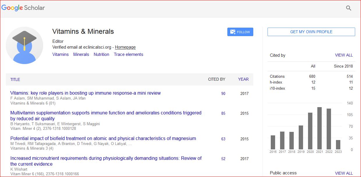
Vitamins & Minerals received 790 citations as per Google Scholar report

 Spanish
  Chinese
  Russian
  German
  French
  Japanese
  Portuguese
  Hindi
Hindi 