Research Article
Pages: 0 - 0Linda S Powers, Walther R. Ellis and Christopher R. Lloyd
Currently, no methods exist for the real-time detection and quantification of microbes in the environment or for the detection and identification of pathogenic organisms in clinical specimens. We have developed technologies which overcome these limitations and provide detection limits as low as a ten microbial cells per cm2 on abiotic surfaces, and per mL in fluids. The detection and quantification of microbes [total microbial load] is based on the intrinsic fluorescence of microbial metabolites and protein cofactors, and provides an estimate of the total microbial load as well as the relative distribution of live cells, dead cells, and endospores. Unlike existing methods, no additional reagents or sample contact is needed. This technology has been applied to the in-situ measurements of two sub-glacial microbial communities at sites in the Svalbard Archipelago, Norway, and to the efficacy of disinfection of contact lenses. In the rapid spread of a life-threatening infection, early diagnosis is of great importance. In such situations, pathogen counts will be very low, which also presents a significant challenge to diagnostic methods. We have developed a point of care disposable diagnostic based on the en masse capture of blood-borne microbes from 1 mL of fresh whole blood with surface-tethered, small molecule ligands. Quantification is based on the intrinsic fluorescence of captured cells.
Research Article
Pages: 0 - 0Sapna Jain, Shree R Singh, Daniel W Horn, Virginia A Davis, Manoj Kumar Ram and Shreekumar Pillai
With increasing reports on bioterrorism and other bio-threats, rapid and real time detection methods for various pathogens are warranted. Attempts have been made to improve electrochemical biosensor performance by incorporating Carbon Nanotubes (CNTs). The high surface area of CNTs allows both immobilization of antibodies and electrochemical measurements. Salmonella monoclonal antibodies were covalently attached onto CNTs by using diimide activated imidation coupling. CNTs functionalized with antibodies were immobilized onto a glassy carbon electrode and the presence of pathogen was detected by studying the changes in charge transfer resistance and impedance, before and after the formation of antigen-antibody complex. CNTs behave as molecular wires allowing electrical communication between the underlying electrode and the conjugated antigen-antibody complex. Nyquist plots and cyclic voltammograms were studied and comparisons have been made between glassy carbon electrodes as working-electrode by itself, electrodes immobilized with antibodies and after the formation of antigen-antibody complex. Cyclic voltammeter experiments had a potential scan rate of 100 mVs-1, step height of 1.0 mV and applied potential from -1.0 V to 1.0 V. The electrochemical impedance experiments applied a frequency range of 100 kHz -100 mHz with an AC sine wave amplitude of 10 mV. Amplification in the current density was observed for CNTs immobilized on the electrode surface and decrease in current density and increased impedance was observed after the antigens bound specific antibodies. Enzyme-Linked Immune Sorbent Assay (ELISA) was done to determine the titer of the antibodies and their sensitivity at different dilutions for antigen detection. This technique could be an effective way to sense the formation of antigen-antibody complexes, with the potential to make the detection process rapid as compared to conventional pathogen detection methods.
Research Article
Pages: 0 - 0Jerrie V. Fairbanks, Linda S Powers, Xiang Zhang, Andrew Duncan and Xavier Ramus
High speed data acquisition architecture is implemented as part of a time-resolved fluorescence detection instrument to directly measure the time course of fluorescent decay. The architecture is implemented using a very fast dynode chain photomultiplier tube and associated gating circuitry, a broad spectrum light emitting diode excitation source, very wide band electronics for amplification and filtering, and a high speed digital oscilloscope. The fluorescence decay of tris (2,2´-bipyridyl) ruthenium (II) is measured and the lifetime measurement is compared with that using other reported methods. The system’s architecture is thereby validated for data acquisition of broadband signals including transient fluorescent recording.
Research Article
Pages: 0 - 0Yaqub Mahnashi and Hussain Alzaher
DOI:
DOI: 10.4172/2155-6210.S11-004
Low frequency continuous time filters are essential analog blocks for biomedical applications. Integrating such filters having large time constants is difficult as it requires large component values. A novel approach to scale down the pole frequency is presented. A 5-bit reduction in the cut off frequency is achieved. This is made possible through adding a passive resistor in the forward path of the op-amp based integrator introducing a difference term of the pole frequency. Also, the filter topology is modified to avoid changing the quality factor. As an example, a 2nd order low pass filter is designed and simulated. Simulation results show that the pole frequency is scaled down from 1.43 MHz to 4.97 kHz while maintaining tuning of 30% around the nominal value by controlling only one resistor.
Research Article
Pages: 0 - 0Longyan Chen and Jin Zhang
In this paper, bioconjugated Magnetic Nanoparticles (MNPs) are developed for rapid capture gram-positive bacterium Staphyloccocus aureus (S. aureus). The MNPs was synthesized through a two-step sol-gel process, followed a bioconjugation of gentamicin (Gm), an aminoglycoside antibiotic, via the linker, glutaraldehyde. The average diameter of the magnetic core is 18 ± 3 nm and the thickness of shell is around 5 ± 3 nm. The XRD results indicate that core-shell MNPs consist of magnetic core, Fe3O4, and silica (SiO2) shell. In addition, the core-shell MNPs show the ferromagnetic properties, whereas the monodipersed Iron Oxide Magnetic Nanoparticles (IONPs), which were produced in the first step, show the typical superparamagnetic properties with a blocking temperature (TB) at 115 K. The interactions between S. aureus and core-shell MNPs with and without Gm have been further investigated by using a Transmission Electron Microscopy (TEM). Our results demonstrate that the diluted S. aureus
with the concentration as low as 0.5 ×10³ CFU/mL can be separated from the solution by the core-shell MNPs in one minute.
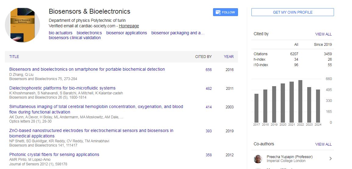
Biosensors & Bioelectronics received 6207 citations as per Google Scholar report

 Spanish
  Chinese
  Russian
  German
  French
  Japanese
  Portuguese
  Hindi
Hindi 