Olena Y. Palyvoda
Scientific Tracks Abstracts: J Biosens Bioelectron
Cancer is recognized as a multistep process involving multiple genomic and epigenomic alterations that occur in multiple phases. The complexity of cancer requires multivariate assays and accurate diagnosis, prognosis and treatment monitoring. These processes are often slow, complex, labor intensive and with sub-optimal sensitivity. Imaging studies such as CT, MRI, radiography and ultrasonography often used to support the diagnosis and stage the tumor. However, despite advances in imaging techniques, there are a number of limitations in sensitivity leading to an inappropriately high rate of misdiagnosed cancers, and an intrinsic inability to prove that a suspicious abnormality is benign or malignant. This has led to the investigation of alternative imaging modalities, such as Raman spectroscopy for early non-invasive detection and diagnosis of different types of cancer. Raman spectroscopy uses visible or near-infrared light to measure a spectrum of vibrational bonds in seconds. Cancer disease leads to chemical and structural changes in tissue that change the vibrational spectra, and that can be used as markers of the disease. Moreover the technique has several advantages over biopsy analysis, including in vivo monitoring, higher sensitivity, easier use and an overall more accurate correlation between cell numbers detected and tumor growth and can be applied to a wide variety of sample morphologies such as thin sections, native tissue, soft tissue, hard tissue and body fluids. For ten years we have used Raman spectroscopy for the diagnosis of malignancy in children. We now propose to extend this work to other difficult areas where a rapid and accurate determination is not yet available such as early diagnosis and treatment outcomes of retinoblastoma and ocular malignancies. We will discuss the label-free optical spectroscopy techniques which are able to non-invasively measure the biochemistry in biological systems and their benefits and limitations, with particular emphasis on applications in biomedicine�both in vivo (using fiber endoscopes) and in vitro (in optical microscopes).
Olena Palyvoda has been an Assistant Professor with the Smart Sensors and Integrated Microsystems (SSIM) group in the Electrical and Computer Engineering Department at Wayne State University. Her areas cover a wide range of fields including: cell imaging, cell-surface biochemistry, surface science, self-assembly, micro- and nanotechnology and development of biomedical microsystems and BioMEMS systems. She also has expertise in molecular biology especially in the mechanisms of the assembly of signaling molecules into complex signalling networks and radiobiology with the identification of radiosensitivity-associated markers that could predict human normal tissue and tumor radioresponse. She is author or co-author of more than 60 published papers and 20 articles in peer-reviewed scientific journals.
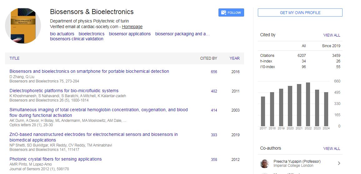
Biosensors & Bioelectronics received 6207 citations as per Google Scholar report

 Spanish
  Chinese
  Russian
  German
  French
  Japanese
  Portuguese
  Hindi
Hindi 