Mohammad RN Avanaki
Accepted Abstracts: J Biom Biostat
Optical imaging is becoming themethod of choice for applications where high-resolution images are required noninvasively. Optical imaging technologies are capable of representing the internal structure of a sample across a range of spatial scales, and that has made them favored tools in biomedical research studies.In the first part of this talk, the authorwill explain the principle of one of the high-resolution optical imaging modalities, optical coherence tomography (OCT), and how it can help in biomedical studies. Just like every other imaging system, OCT systems have limitations. Three separate limitations will be discussed. First, there is speckle noise due to the use of a broadband illumination source. Second, there is intensity decay due to tissue absorption. And finally, there are aberrations and blurriness due to point spread function (PSF) distortion. The author will then discuss a number of algorithms devised to reduce the impact of these limitations. Moreover, how by using the enhanced Huygens-Fresnel (eHF)light propagation theorem, one can extract optical properties from a specific region in an OCT image will be explained. This ability is important in automating diseasediagnostics and adding more data to the morphological information that the OCT has already provided. As concrete evidence, the results of using this method to differentiate basaloid and healthy tissues will also be demonstrated.In the second part of this talk, the author will look into another high-resolution imaging modality, photoacoustic tomography. As opposed to OCT, where the attenuation of the backscattered light reaching the detector limits the penetration depth, photoacoustic tomography uses optical excitation and acoustic detection, and dramatically increases the penetration depth. The author will explain howhedeveloped a functional connectivity photoacoustic tomography (fcPAT)system, which allowed, for the first time, non-invasive imaging of resting-state functional connectivity (RSFC) in the mouse brain, with a large field of view and high spatial resolution. This neuroimaging technique can easily be applied to mice, the most widely used model species for human brain disease studies. Results that showfcPAT is a promising, noninvasive technique for functional imaging of the mouse brain will be demonstrated. Due to the low cost of fcPAT compared with current functional imaging modalities such as functional Magnetic Resonance Imaging (fMRI), it is expectedthat fcPAT will enable many laboratories that previously did not consider functional neuroimaging to contribute further to ongoing studies of brain disease.
Mohammad R N Avanaki received his PhD in Medical Image Computing from the University of Kent in the United Kingdom. His Bachelor?s and Master?s degrees with honors are in Electronics Engineering. He also holds a Post Graduate Certificate for Higher Education research and teaching from the University of Kent. As a faculty member, he was awarded the ?Best Lecturer Award? at Azad University of Karaj for excellence in teaching. He has published more than 27 first-authored peer-reviewed articles in journals such as PNAS, IEEE Photonics Technology Letters, Applied Optics, and Modern Optics. He has also presented at conferences across the globe, such as MICCAI, MIUA, MVIP, and Photonics West, and has received several best paper awards. He is a member of the technical program committee of IEEE ICIP 2014 and MICCAI 2014. He has peer-reviewed for PNAS, PLOS ONE, MICCAI, Optics Letters, Optics Express, and Journal of Biomedical Optics. He is currently a Research Associate in the Optical Imaging Laboratory at the Washington University in Saint Louis. His research focuses on using highresolution medical imaging modalities, including optical coherence tomography and photoacoustic technology, in biomedical applications
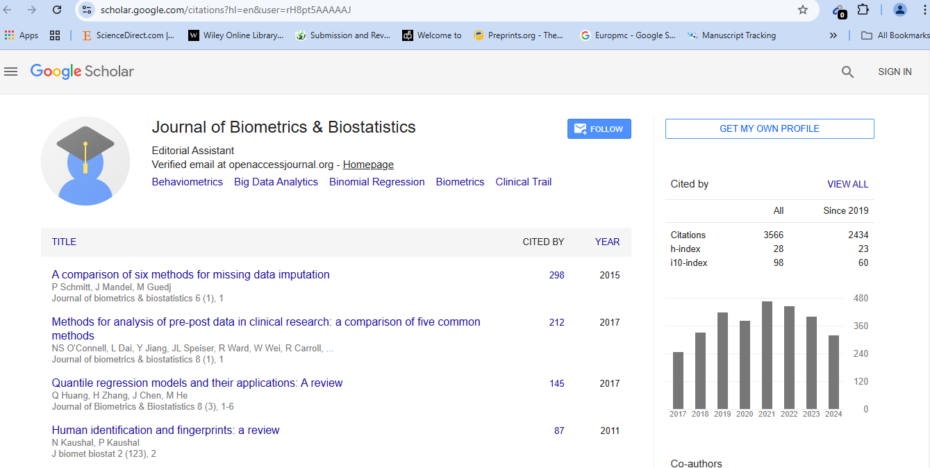
Journal of Biometrics & Biostatistics received 3496 citations as per Google Scholar report

 Spanish
  Chinese
  Russian
  German
  French
  Japanese
  Portuguese
  Hindi
Hindi 