Case Report - (2026) Volume 10, Issue 1
Received: 05-Jan-2026, Manuscript No. cmcr-26-180431;
Editor assigned: 07-Jan-2026, Pre QC No. P-180431;
Reviewed: 20-Jan-2026, QC No. Q-180431;
Revised: 23-Jan-2025, Manuscript No. R-180431;
Published:
03-Feb-2026
, DOI: 10.5281/zenodo.18410213
Citation: Calabrez, Jullia Greque and Victoria Caroline Danielli da Silva. âUterine Arteriovenous Malformation as a Cause of Postpartum Hemorrhage: A Case Report.â Clin Med Case Rep 11 (2026): 412.
Copyright: © 2026 Calabrez JG, et al. This is an open-access article distributed under the terms of the Creative Commons Attribution License, which permits unrestricted use, distribution and reproduction in any medium, provided the original author and source are credited.
Introduction: Arteriovenous Malformation (AVM) is a rare vascular abnormality characterized by a direct connection between arteries and veins without an intervening capillary network, resulting in increased circulatory volume and a risk of severe complications. In the gynecological context, uterine AVM is uncommon but clinically significant due to its association with severe and difficult-to-control hemorrhage. Objective: To describe the case of a primiparous patient who was diagnosed with a uterine arteriovenous malformation following cesarean delivery, highlighting the clinical findings and the management approach adopted during follow-up. Methods: This descriptive study was conducted as a clinical case report, based on a retrospective analysis of clinical, laboratory, and imaging data from a patient diagnosed with AVM. Data were collected through a systematic review of the electronic medical records of Hospital Santa Casa de Misericórdia de Vitória (HSCMV), ensuring compliance with ethical principles and data confidentiality. Expected results: This case report is expected to contribute to the expansion of knowledge regarding uterine AVM. The findings are anticipated to emphasize the importance of early diagnosis, even in cases with nonspecific clinical manifestations, as well as to highlight the challenges associated with therapeutic management. Consequently, this report aims to raise awareness among healthcare professionals regarding subtle clinical signs of the condition and to promote more accurate and appropriate clinical decision-making in relevant settings.
Uterine hemorrhage • Arteriovenous fistula • Postpartum hemorrhage • Hysterectomy
CEP: Research Ethics Committee; CNS: National Health Council; AVM: Arteriovenous Malformations; ICF: Informed Consent Form; HSCMV: Santa Casa de Misericórdia de Vitória Hospital
Arteriovenous Malformation (AVM) is a rare vascular abnormality characterized by a direct connection between arteries and veins without an intervening capillary bed. This alteration results in disorganized blood flow, leading to increased circulatory volume and potentially causing severe complications and significant hemodynamic consequences.
Acquired AVMs are abnormal communications between intramural branches of the uterine artery and the myometrial venous plexus, located within the myometrium and endometrium [1]. Clinically, AVMs may present in a wide spectrum, ranging from nonspecific symptoms to severe manifestations such as hemorrhage, epileptic seizures, and neurological deficits, depending on the location of the lesion. As emphasized by Siqueira EC, et al. [2]. “AVMs consist of a tangled network of dilated blood vessels in which arterial blood is shunted directly into the venous system without passing through the capillary network, resulting in hemodynamic overload and an increased risk of rupture.”
In the gynecological context, AVMs most commonly occur in the uterus. Although uncommon, they are of high clinical relevance due to their frequent association with severe and difficult-to-manage uterine bleeding. These lesions may be congenital or acquired and are usually related to traumatic events or surgical procedures such as curettage, cesarean section, or myomectomy. “uterine arteriovenous malformations, although rare, should be included in the differential diagnosis of abnormal uterine bleeding, particularly in women of reproductive age, with Doppler ultrasonography being the first-line imaging modality for investigation.” In addition, the literature highlights that magnetic resonance imaging and angiography play a fundamental role in confirming the diagnosis and guiding therapeutic management.
Therefore, early and accurate diagnosis of AVMs is essential not only for selecting the most appropriate treatment but also for preventing potentially life-threatening complications. As reinforced by Vilos, et al. selective arterial embolization has become the treatment of choice in symptomatic cases, as it “effectively controls hemorrhage while preserving reproductive function.” Thus, understanding the clinical and diagnostic aspects of AVMs, particularly uterine AVMs, is crucial to ensure patient survival and health, reduce morbidity and mortality, and promote better clinical outcomes.
Data collection was performed through a retrospective analysis of the patient’s medical records, complemented by a review of laboratory tests, imaging studies, and clinical follow-up notes. Inclusion criteria were based on diagnostic confirmation and complete therapeutic follow-up. The study adhered to ethical principles and informed consent was obtained from the patient for the publication of data, ensuring anonymity and confidentiality of the information. The adopted methodology allowed for an accurate reconstruction of the case and a critical analysis of the clinical events.
A 23-year-old primigravida, mixed-race female with no prior comorbidities underwent prenatal care in the public health system and was classified as low-risk, with no significant gestational complications. She went into labor and subsequently underwent cesarean delivery at another hospital due to acute fetal distress with fetal bradycardia following a diagnosis of arrest of labor progression and failed vacuum-assisted delivery. The surgical procedure was technically challenging due to fetal macrosomia and was complicated by uterine atony and significant hemorrhage, which were managed with uterine massage, administration of uterotonic agents, application of a B-Lynch hemostatic suture, and blood transfusion. The patient required hemotransfusion and treatment for puerperal endometritis, for which antibiotic therapy with gentamicin and clindamycin was administered. No additional procedures were necessary. After clinical stabilization, the patient was discharged from the hospital.
After hospital discharge, the patient sought care at a referral hospital for gynecology and obstetrics with specialized emergency services. She was admitted on the 58th day postpartum, reporting heavy vaginal bleeding associated with cramp-like abdominal pain. On initial physical examination, she was conscious and oriented, pale (+/4+), hydrated, with a blood pressure of 110/80 mmHg, an appropriately involuting uterus, and moderate vaginal bleeding. During the initial assessment, symptomatic medication was prescribed and reassessment was recommended. After initial care, the patient experienced an episode of syncope accompanied by profuse vaginal hemorrhage. Vital signs at that time showed a blood pressure of 98/54 mmHg and a heart rate of 88 bpm. Given the severity of bleeding, signs of anemia on inspection, and borderline blood pressure with altered heart rate, the patient was admitted to the institution for the first time for clinical stabilization and diagnostic investigation.
Initial laboratory tests revealed the following results: hemoglobin 8.9 g/dL, hematocrit 26.5%, INR 1.17, aPTT 26 s, white blood cell count 7,590/mm³, platelet count 245,000/mm³, total bilirubin 0.5 mg/dL, and fibrinogen 229 mg/dL. In view of the anemia and active bleeding, treatment was initiated with intravenous tranexamic acid and oral iron supplementation (ferrous sulfate), and a Transvaginal Ultrasound (TVUS) was requested.
On the following day, repeat laboratory tests were requested and revealed hemoglobin of 7.1 g/dL, hematocrit of 21.4%, platelet count of 233,000/mm³, and a normal coagulation profile. Due to the significant decline in hemoglobin levels, transfusion of one unit of packed red blood cells was performed.
Transvaginal Ultrasonography (TVUS) demonstrated a uterus in anteverted and anteflexed position, centrally located, with a volume of 102.1 cm³, regular contours, and well-defined margins. In the isthmic region and left posterolateral uterine wall, a solid nodular lesion was identified, echogenic relative to the myometrium, heterogeneous in appearance, with myofasciculation signs and the presence of dilated vessels exhibiting intense arterial flow directed toward the center of the lesion, possibly originating from the hypogastric (internal iliac) vessels. The endometrium measured 2.9 mm in thickness, with a homogeneous and well-defined surface. No free fluid or masses were observed in the posterior cul-de-sac (Figures 1-3).
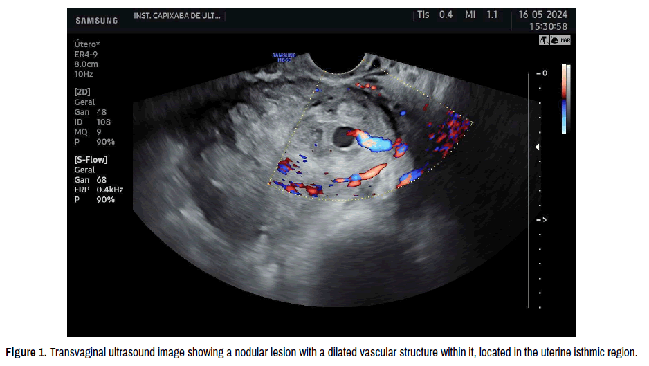
Figure 1. Transvaginal ultrasound image showing a nodular lesion with a dilated vascular structure within it, located in the uterine isthmic region.
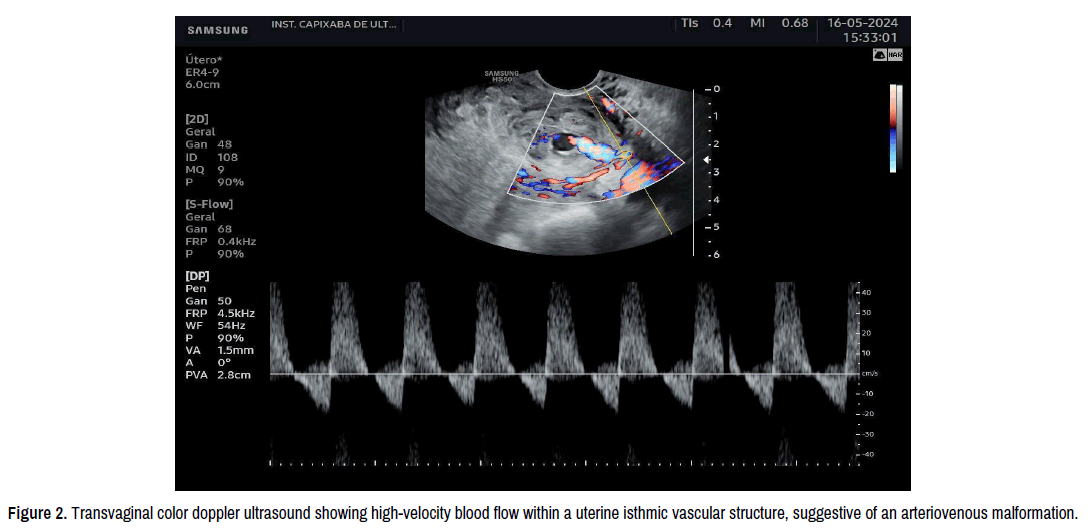
Figure 2. Transvaginal color doppler ultrasound showing high-velocity blood flow within a uterine isthmic vascular structure, suggestive of an arteriovenous malformation.
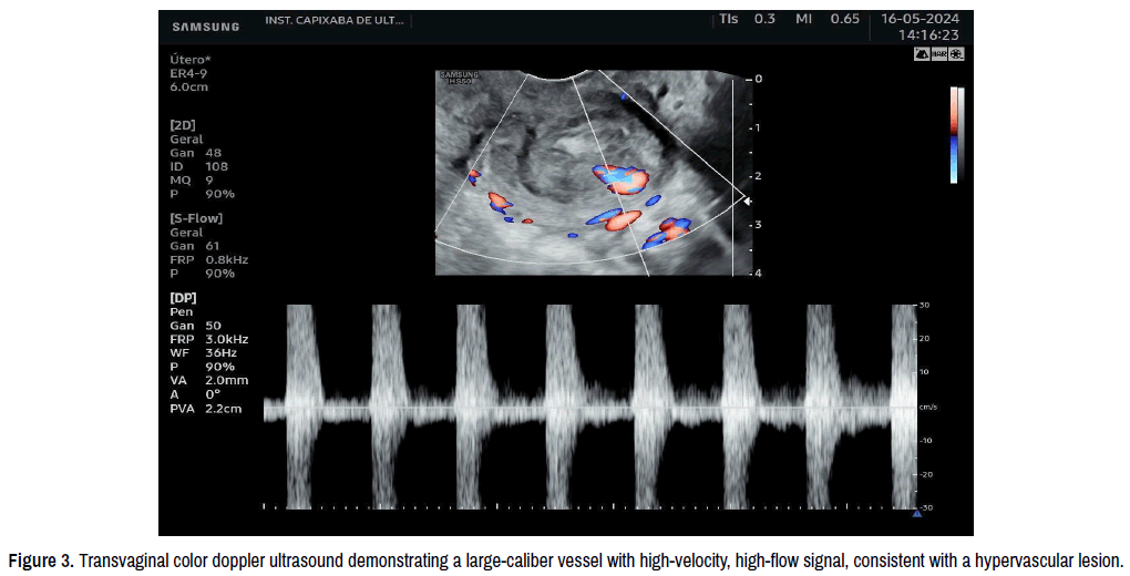
Figure 3. Transvaginal color doppler ultrasound demonstrating a large-caliber vessel with high-velocity, high-flow signal, consistent with a hypervascular lesion.
Following the transvaginal ultrasound findings, the patient was kept hospitalized for bleeding surveillance, with laboratory testing every 48 hours to monitor hematimetric parameters and ensure clinical stabilization. A repeat complete blood count performed after blood transfusion showed hemoglobin of 7.9 g/dL, hematocrit of 26.8%, and platelet count of 206,000/mm³. The patient continued to have vaginal bleeding, which was partially controlled with intravenous tranexamic acid. Subsequently, a repeat transvaginal ultrasound was requested to assess the evolution of the uterine lesion. The follow-up transvaginal ultrasound revealed, in the isthmic region of the left posterolateral uterine wall, a solid nodular lesion, echogenic relative to the myometrium, heterogeneous, with myofasciculation signs, and a large vessel with intense arterial flow reaching the central portion of the lesion, appearing to be a branch of the uterine artery. The endometrial thickness was 2.9 mm, and the ovaries showed no abnormalities. Figure 4 illustrates the transvaginal ultrasound findings.
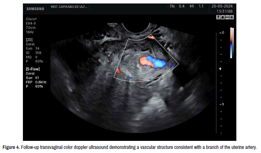
Figure 4. Follow-up transvaginal color doppler ultrasound demonstrating a vascular structure consistent with a branch of the uterine artery.
After 24 hours, the patient underwent a new clinical reassessment and follow-up imaging. She was considered suitable for hospital discharge with outpatient follow-up, given her clinical stability and complete resolution of vaginal bleeding. She was advised to return for weekly follow-up imaging or immediately in the event of any new clinical deterioration. Subsequently, a follow-up outpatient ultrasound was performed as recommended at discharge (Figure 5). The patient remained asymptomatic, with no vaginal bleeding, and imaging demonstrated persistence of the lesion in the uterine isthmic region. As previously instructed to return in the presence of warning signs, three days after the last ultrasound examination, the patient presented to the emergency department reporting heavy vaginal bleeding with clot passage, associated with weakness and tremors. On physical examination, she remained pale, with no signs of peritoneal irritation. Gynecological examination revealed profuse vaginal bleeding with clots. In view of this presentation, hospital admission was indicated (Figure 6).
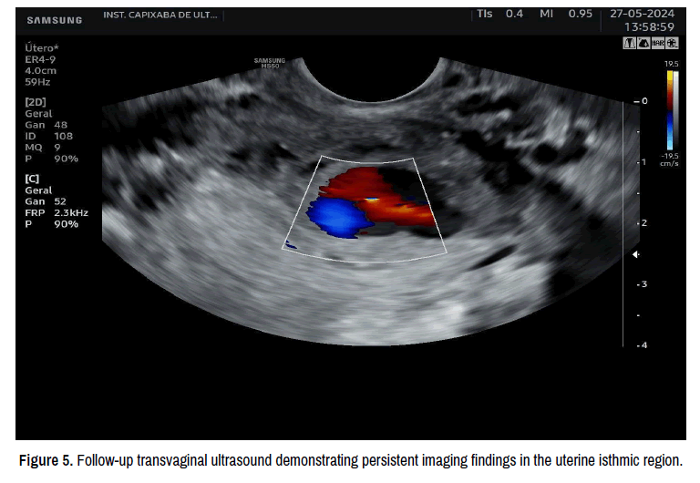
Figure 5. Follow-up transvaginal ultrasound demonstrating persistent imaging findings in the uterine isthmic region.
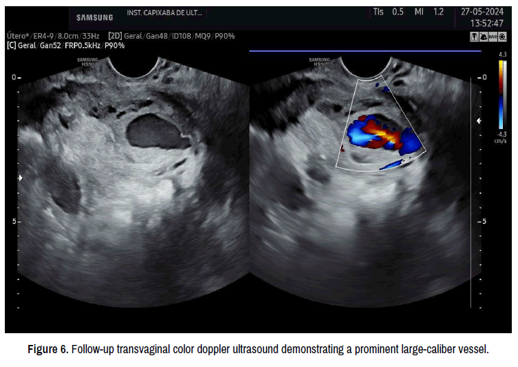
Figure 6. Follow-up transvaginal color doppler ultrasound demonstrating a prominent large-caliber vessel.
During hospitalization, the patient was kept under ward observation, and within the first 24 hours she experienced worsening vaginal bleeding, although vital signs initially remained stable. Repeat laboratory tests showed hemoglobin of 9.6 g/dL, hematocrit of 29.8%, platelet count of 232,000/mm³, negative beta-human chorionic gonadotropin (β-hCG), and INR of 1.09. Twenty-four hours after the episode of intense vaginal bleeding, the patient developed hemorrhagic hypovolemic shock, characterized by tachycardia (heart rate 140 bpm), hypotension (blood pressure 90/60 mmHg), and signs of severe hemodynamic instability. Initial stabilization measures were promptly instituted, including blood transfusion and aggressive volume resuscitation. Given the critical condition and the failure of conservative bleeding control, a total abdominal hysterectomy was indicated (Figure 7). During surgery, the uterus was found to be friable, with the presence of sutures consistent with the B-Lynch technique, and a vascular lesion in the region of the left uterine artery, associated with a blood clot. As illustrated in Figure 8, a bulging lesion in the topography of the left uterine artery was identified, the rupture of which exposed a thrombotic formation.
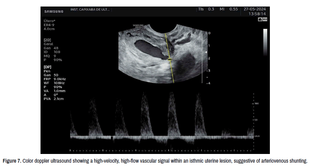
Figure 7. Color doppler ultrasound showing a high-velocity, high-flow vascular signal within an isthmic uterine lesion, suggestive of arteriovenous shunting.
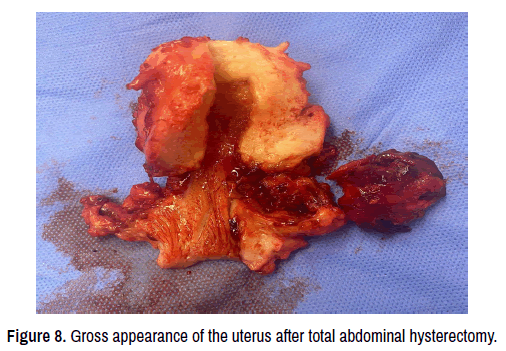
Figure 8. Gross appearance of the uterus after total abdominal hysterectomy.
The adnexa were preserved, and the surgical specimen was sent for histopathological examination. Gross (Figures 9 and 10) and microscopic analyses (Figures 11-14) were performed, confirming the presence of a uterine arteriovenous malformation with isthmic rupture, in addition to an angioleiomyoma, proliferative endometrium, tubal congestion, and no evidence of malignant neoplasia. The postoperative course was uneventful, with no major complications. The patient showed progressive clinical improvement and was discharged from the hospital in good general condition, with appropriate clinical guidance, symptomatic medication, and scheduled outpatient follow-up.
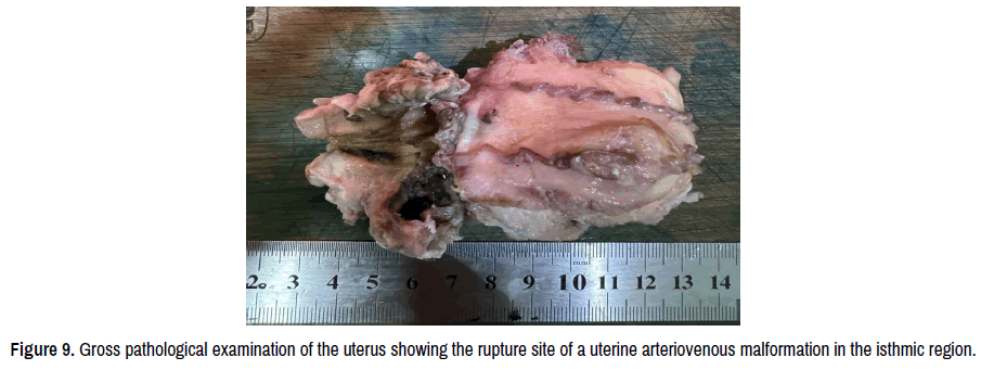
Figure 9. Gross pathological examination of the uterus showing the rupture site of a uterine arteriovenous malformation in the isthmic region.
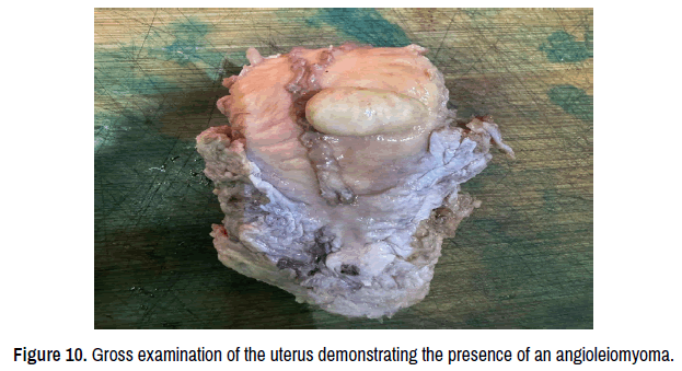
Figure 10. Gross examination of the uterus demonstrating the presence of an angioleiomyoma.
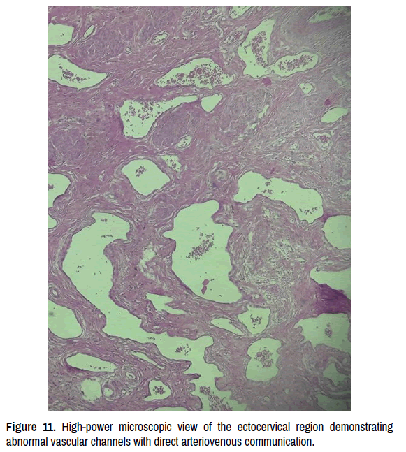
Figure 11. High-power microscopic view of the ectocervical region demonstrating abnormal vascular channels with direct arteriovenous communication.
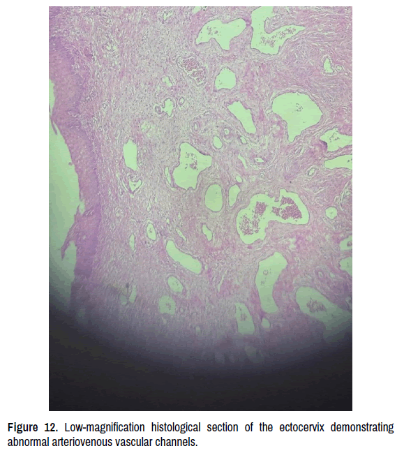
Figure 12. Low-magnification histological section of the ectocervix demonstrating abnormal arteriovenous vascular channels.
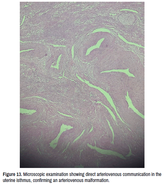
Figure 13. Microscopic examination showing direct arteriovenous communication in the uterine isthmus, confirming an arteriovenous malformation.
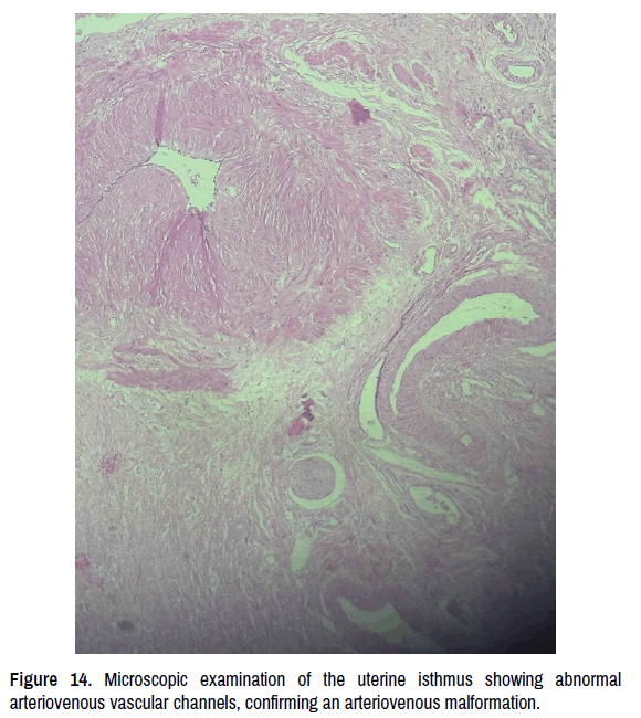
Figure 14. Microscopic examination of the uterine isthmus showing abnormal arteriovenous vascular channels, confirming an arteriovenous malformation.
Uterine Arteriovenous Malformations (AVMs) are rare and frequently underdiagnosed conditions, representing a clinical challenge due to their variable presentation and potential severity. Uterine AVMs typically present with sudden and heavy vaginal bleeding [3]. They may be congenital, resulting from abnormalities in embryonic vascular development, or acquired, most commonly associated with intrauterine procedures such as cesarean section, curettage, myomectomy, or traumatic deliveries [4, 5]. In the present case, the uterine AVM presented as an acquired lesion, likely secondary to surgical trauma and vascular repair processes following cesarean delivery, which itself is a traumatic procedure. The classic clinical presentation involves persistent or recurrent abnormal uterine bleeding, often heavy and difficult to control, which may occur weeks or months after the precipitating event. This characteristic may delay diagnosis, particularly in the postpartum period, when bleeding is expected and may mask the severity of the condition. As emphasized by Amiri and Hamidi, clinical suspicion should be heightened in cases of disproportionate or treatment-refractory hemorrhage, especially in patients with a recent history of uterine manipulation.
Imaging studies play a fundamental role in the diagnosis of AVMs. Pelvic ultrasonography with Doppler is considered the initial diagnostic modality of choice, as it allows identification of high-velocity arterial flow, turbulent patterns, and ectatic vessels, which are suggestive of this vascular anomaly. As noted by Farias MS, et al. [1], Doppler ultrasonography is a noninvasive and widely available method, and awareness and recognition of this clinical entity are essential despite its rarity. Magnetic resonance imaging and angiography, in turn, provide essential anatomical and hemodynamic detail, assisting in therapeutic planning and confirming the diagnosis. In this patient, ultrasonography was crucial in raising diagnostic suspicion and, when the patient progressed to hemorrhagic shock, in guiding the surgical management strategy.
Therapeutic management depends directly on the clinical severity and the patient’s hemodynamic stability. In asymptomatic cases or those with mild and controllable bleeding, conservative management with clinical follow-up may be considered. However, in symptomatic cases, selective uterine artery embolization is regarded as the first-line treatment, with high success rates and preservation of fertility. Nevertheless, in the presence of severe hemodynamic instability or failure of conservative measures as observed in the present case, total hysterectomy remains the definitive and life-saving treatment [2].
The concomitant presence of an angioleiomyoma on histopathological examination highlights the importance of thorough pathological evaluation. Although rare, this finding may indicate the coexistence of benign lesions and severe vascular anomalies, potentially influencing prognosis and management Torres MA, et al. [6]. Thus, a multidisciplinary approach involving gynecology, radiology, and pathology is essential for optimal patient care. This case report underscores the need for high clinical suspicion and a rapid, accurate diagnostic approach in cases of severe postpartum uterine hemorrhage, particularly when associated with prior obstetric procedures. Early diagnosis and timely intervention are crucial for patient survival and for reducing serious complications, including hypovolemic shock and maternal death [7-10].
The present case report highlights the importance of considering uterine Arteriovenous Malformations (AVMs) in the differential diagnosis of acute gynecological hemorrhage, particularly in young women of reproductive age with a recent history of obstetric procedures. “the approach to late postpartum hemorrhage is always challenging; initial assessment and stabilization of the patient are essential, and therapeutic interventions should not be undertaken before identifying the underlying cause, as this may worsen the prognosis.” The rapid and potentially fatal clinical course, marked by the risk of hypovolemic shock and hemodynamic instability, underscores the need for early recognition, appropriate imaging investigation, and timely therapeutic decision-making.
Although hysterectomy represents a radical intervention with a significant impact on fertility, in emergency settings and in cases of failure of conservative management, it remains the definitive and life-saving treatment. When feasible, ovarian preservation, as achieved in the present case, may help maintain hormonal function and prevent premature menopause, thereby contributing to better long-term quality of life. Furthermore, although gestation within the patient’s own body is no longer possible due to the absence of the uterus, reproductive options remain available through assisted reproductive techniques, such as in vitro fertilization with gestational surrogacy. While not universally accessible, these alternatives may still represent a possibility for future parenthood. This case also emphasizes the relevance of a multidisciplinary approach and close clinical surveillance to minimize complications and ensure patient survival, highlighting the need for greater awareness of this rare but high-impact condition in gynecological practice.
Google Scholar, Cross Ref, Indexed at
Google Scholar, Cross Ref, Indexed at
Google Scholar, Cross Ref, Indexed at
Google Scholar, Cross Ref, Indexed at
Google Scholar, Cross Ref, Indexed at
Google Scholar, Cross Ref, Indexed at
Google Scholar, Cross Ref, Indexed at
Clinical and Medical Case Reports received 53 citations as per Google Scholar report