Commentary - (2022) Volume 13, Issue 3
Received: 17-Feb-2022, Manuscript No. jbsbe-22-66139;
Editor assigned: 20-Feb-2022, Pre QC No. P-66139;
Reviewed: 25-Feb-2022, QC No. Q-66139;
Revised: 04-Mar-2022, Manuscript No. R-66139;
Published:
08-Mar-2022
, DOI: 10.37421/2155-6210.22.13.319
Citation: Lin, Yuehe. "Uses of ELISA in Cancer Biomarker Detection." J Biosens Bioelectron 13 (2022): 319.
Copyright: © 2022 Lin Y. This is an open-access article distributed under the terms of the Creative Commons Attribution License, which permits unrestricted use, distribution, and reproduction in any medium, provided the original author and source are credited.
Cancer is one of the leading causes of death worldwide. Many variables, including as exposure to cancer-causing chemicals, radiation, infections, genetic alterations, and so on, can disturb cells and cause them to change and proliferate, resulting in the development of cancer in various regions of the body. It is not suggested to diagnose it purely on visual symptoms since such symptoms emerge in later stages of cancer when there are no effective treatments. As a result, it is recommended that cancer patients be diagnosed early on, when therapy is still effective, in order to increase their chances of survival. Researchers have proposed using proteins and oligonucleotides secreted in the body during the early stages of cancer and not present in the blood to accomplish early stage detection [1].
Biomarkers are chemicals that are released by many forms of malignancies, and their detection and quantification can give extremely useful information about the cancer type and stage. As a result, developing systems that are simple, low-cost, and capable of providing sensitive and specific estimate of such biomarkers is critical. Furthermore, to improve diagnostic accuracy, it is essential to discover and test panels of numerous biomarkers, taking into consideration demographic and cancer stage variability, as well as low levels of biomarkers in early stages of cancer. Furthermore, these biomarkers should be detected in a non-invasive or minimally invasive way with good selectivity, sensitivity, and no false positives or negatives [2].
Biomarkers are used in many cancer detection technologies, including enzyme-linked immunosorbent assay (ELISA), western blotting, optical, electrochemical, fluorescent, or radio immunosensor-based systems, and their estimated levels are connected to cancer stage and guide cancer therapy. Researchers have found many possible biomarkers unique to individual tumours and connected to the bio-mechanics of cancer cells as a result of breakthroughs in cancer biology and immunology. Optical sandwich ELISAbased biomolecule detection is still widely used in clinical practise and is widely regarded as the gold standard approach. In a technique known as immunoassay, antibodies are employed to specifically identify and quantify the target antigen/biomarker; sensors utilised in these assays are known as immunosensors [3].
Traditional optical ELISA is often performed on 96-well plates in the medical diagnostics business. For required analytes testing and estimation, suppliers supply reagent kits and 96 well plates. A primary antibody is deposited onto the wells of the plate through physical adsorption and then blocked to avoid non-specific binding in such kits. Operating instructions are also included in the kits. To produce an antibody–antigen complex, an antigen sample is first incubated in the well with primary antibodies for the appropriate period. Plates are typically cleaned with wash buffer given by the kit vendor after incubation. To make an antibody-antigen-antibody sandwich, the antigenantibody complex is washed and incubated with an enzyme-tagged detection antibody, following the incubation period [3].
During incubation, the enzymatic reaction causes the indicator dye to change colour, which may be measured using an optical reader to determine the absorbance value. The analyte concentration is determined by comparing the absorbance value to the standard solution calibration. The entire testing technique is time-consuming, and analyte estimation frequently necessitates the use of a costly optical reader. The employment of a sandwich approach, on the other hand, results in an amplified response and hence a greater detection range. In summary, optical ELISA gives quantitative data that is highly repeatable, sensitive, and specific, making it a useful biotechnological tool in scientific study and clinical diagnostics. Optical ELISA, on the other hand, has tedious/laborious processes, requires centralised laboratory equipment, and requires a relatively large sample volume [4].
Furthermore, the detection limit of traditional ELISA is just below the nanomolar concentration level, which is insufficient to achieve the clinical threshold of many protein biomarkers, particularly in the early stages of illness. Electrochemical tests have showed promise in dealing with these problems. Electrochemical assays have the advantages of being simple to perform, portable, low volume, and rapid measurements. However, electrochemical assays have not had as much success as optical ELISA in 96 wells accomplishing huge multiplexing concurrently. Electrochemical ELISA has shown promise among electrochemical assays, as it combines the advantages of optical ELISA, such as sensitivity and specificity, multiplexing, and quantitative data, with the advantages of an electrochemical assay, such as speed, lower sample volume, and low-cost instrumentation.
Various researchers have suggested novel technologies, such as enhanced sensor surfaces and detecting probes, to reduce the time required and improve the responsiveness and features of classic optical ELISA. Sandwich-based electrochemical ELISAs have also been proposed to reduce cost, simplify testing, and shorten measurement time by combining the specificity of optical ELISA with the advantages of electrochemical measurements to achieve better response and characteristics for desired analyte estimation. Electrochemical ELISA, unlike optical ELISA, employs a potentiostat/galvanostat to assess signal in research laboratories. Though there aren't many commercial electrochemical ELISA-based immunosensors on the market right now, the necessary gear is accessible, and there's a lot of room for such sensors and their commercialization. Furthermore, the simplicity with which essential electronics may be miniaturised opens the door to smaller, simpler, and lower-cost devices for such measurements. In summary, electrochemical immunosensors have been proposed as a viable alternative to optical ELISA for overcoming constraints while keeping the benefits of classical tests. Electrochemical immunosensors based on potential, current, or impedance may give the needed sensitivity in extremely low volume samples at a quicker rate of analysis while also being simple to fabricate, monitor, and mass produce at a cheap cost. With these benefits in mind, researchers have lately concentrated on the development of electrochemical ELISA-based systems that combine the benefits of sandwich assays used in optical ELISA with electrochemical detection.
Electrochemical ELISA benefits from the high sensitivity, low detection limit, simple handling, and easy detection in a downsized format afforded by electrochemical detection, as well as high sensitivity, low detection limit, easy handling, and easy detection in a tiny format. With advances in material and surface chemistry, as well as bio- and nanotechnologies, electrochemical ELISA-based immunosensors have attracted a lot of attention and promise to replace traditional optical ELISA for faster, more sensitive, less expensive, and more reliable detection of cancer biomarkers in early stage diagnosis. The current review discusses several innovative methods for creating and upgrading sandwich-based electrochemical ELISA for cancer biomarker detection that have been published by researchers in the recent 3 to 4 years.
Many of these studies' authors confirmed their methods in vitro using spiked/real samples. There is currently no information on the commercialization of any of these sensors, but these studies may pave the way for better and faster cancer detection at an earlier stage in the near future. There are also a number of excellent reviews for electrochemical immunosensor-based cancer biomarker detection employing nanoelectrodes, arrays, and microfluidics that have been published in the past. Combining the recent developments in sensor surfaces and detection probes presented here with nanoelectrode arrays or microfluidics in the future may improve the prospects of reaching greater sensitivity and detection limits for early stage biomarker assessments [5].
None.
Google Scholar, Crossref, Indexed at
Google Scholar, Crossref, Indexed at
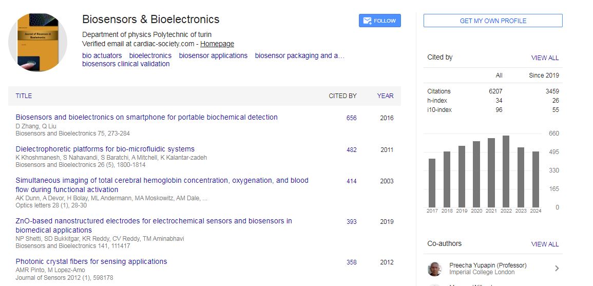
Biosensors & Bioelectronics received 6207 citations as per Google Scholar report