Case Report - (2022) Volume 11, Issue 5
Received: 12-May-2022, Manuscript No. JTM-22-63737;
Editor assigned: 13-May-2022, Pre QC No. P-63737;
Reviewed: 21-May-2022, QC No. Q-63737;
Revised: 31-May-2022, Manuscript No. R-63737;
Published:
06-Jun-2022
, DOI: 10.37421/2167-1222.2022.11.507
Citation: Brigmon, Erika, Ashley Chakales, Derek Lumbard and Matthew Marcus et al. “Tracheal Invasive Mucormycosis after Blunt Trauma: A Case Report.” J Trauma Treat 11 (2022): 507.
Copyright: © 2022 Brigmon E. This is an open-access article distributed under the terms of the Creative Commons Attribution License, which permits unrestricted use, distribution, and reproduction in any medium, provided the original author and source are credited.
Mucormycosis is a rare and highly lethal fungal infection, with a subtle clinical presentation but aggressive spread via angioinvasion. It is uncommon and typically seen in immunocompromised patients but can also be a serious complication after traumatic injuries. Recognition of this disease requires high clinical suspicion and prompt interventions. We report a fatal case of mucormycosis tracheitis in a 23-year-old male after blunt trauma treated at a Level 1 trauma center.
Invasive fungal infection • Mucormycosis • Blunt trauma.
Mucormycosis is a rare and highly lethal angio- invasive fungal infection caused by any fungi in the order Mucorales that leads to thrombosis and subsequent tissue necrosis [1]. It is ubiquitous in the environment, particularly found in decaying vegetation and contaminated soil which makes avoiding exposure virtually impossible [1]. A majority of patients with invasive mucormycosis have some underlying condition, such as diabetes mellitus, hematologic malignancy, trauma, iron overload or a history of a transplant that predisposed them to develop a clinical infection [1,2]. However, cases have been documented in otherwise healthy trauma patients [2] who acquired the infection via direct inoculation of the spores into the injured area [3]. The annual incidence of invasive fungal infections among the civilian population is approximately 0.43 - 1.7 cases per million [4]. Of all cases, 21% to 59% of primary cutaneous mucormycosis are attributed to natural disasters and traumatic injury [4,5] inclusive of motor vehicle collisions, agricultural trauma, and falls [3].
In a 6-year retrospective chart review of 16 patients in Texas, a cluster of mucormycosis cases were noted during the months of February and March when the average temperature rarely exceeded 25ºC [6]. The demographics of invasive fungal infections reflect that of the general trauma population with a male predominance and a mean age between 27 and 48 years old [7]. Here, we report a rare fatal case of mucormycosis tracheitis in a 23-year-old male after blunt trauma in a woodland area in West Texas.
A 23-year-old male with an unknown past medical history presented to a level 1 trauma center after falling approximately 100 ft off a cliff in rural Texas. On admission, he was hemodynamically unstable requiring active whole blood transfusion with a Glasgow Coma Score (GCS) of 3. A liter of hemothorax was evacuated with a right thoracostomy based on chest X-ray findings. He was brought emergently to the operating room for exploration. A right thoracotomy revealed active bleeding from the right inferior posterior mediastinum concerning for an Inferior Vena Cava (IVC) injury. We proceeded to extend to a clamshell thoracotomy and exploratory laparotomy with mobilization of the retro hepatic IVC and partial diaphragm takedown. A partial thickness tear through the longitudinal muscular layer of the esophagus was also identified and repaired. The multidisciplinary team approach was completed with interventional radiology performing an angiogram in which bleeding from the celiac artery branch was identified, and subsequent coil embolization attempted, resultingin unintentional coil deployment into the right hepatic artery. Attempts to recover the coil were halted due to hemodynamic instability and the abdomen was re-entered finding a 1 cm defect at the celiac trunk origin off the aorta that was primarily repaired. The estimated blood loss was 10 L and a total of 24 units of whole blood were given intraoperatively. A temporary abdominal closure of the chest and abdomen was performed, and the patient transferred to the surgical intensive care unit for further resuscitation. He was taken back to the operating room for definitive closure of his chest and abdomen within 24 hours after resuscitation.
His hospital stay was significant for persistent fevers associated with shock liver secondary to ischemia (Figure 1). Broad-spectrum antibiotics were started with no other source identified. On hospital day 6, he was briefly extubated and found to be neurologically intact but required high flow nasal cannula. He was subsequently reintubated less than 12 hours later. After reintubation, a bronchoscopy with bronchioalveolar lavage (BAL) was performed in which mucopurulent secretions were noted. Empiric antibiotic treatment for ventilator associated pneumonia was started. Due to persistent clinical signs concerning for sepsis, a Fungitell test was sent and reported as negative. On hospital stay day 11, he developed redness, warmth and edema over the upper anterior chest wall and continued to be febrile with a worsening leukocytosis. Given his skin findings, his history of prolonged extrication from the side of a cliff and worsening signs of sepsis on broad antibiotics without identified source, suspicion was raised for cutaneous mucormycosis. Liposomal amphotericin B was started empirically, and emergent skin biopsy was completed but was negative for any invasive fungal infection. His Liposomal amphotericin B was continued while waiting for the final fungal culture results. Despite on-going medical treatment, he continued to decompensate and on hospital day 15, a CT scan of his chest showed new mediastinitis (Figures 1 and 2). A bronchoscopy was repeated, this time revealing an approximately 8 cm long area of necrosis at the posterior aspect of the trachea (Figure 3). The next day he went to the operating room for debridement of the mediastinum and primary repair of a tracheal perforation identified intraoperatively. A fresh intraoperative sample was taken for pathology studies which allowed histological confirmation of aseptate hyphae compatible with mucormycosis infection (Figure 4). Systemic posaconazole and amphotericin nebulization were added to his antifungal regimen. Despite the broad antifungal coverage, he continued to deteriorate with massive air leaks from the chest tubes, increasing vasopressor requirement, multi-organ failure, severe acidosis requiring Continuous Renal Replacement Therapy (CRRT), and ST segment elevations on electrocardiogram (ECG) concerning for pericarditis. Repeat bronchoscopy confirmed progression of the fungal invasion into the bronchi with extensive tracheal necrosis and perforation. These findings were deemed incompatible with life, and the patient was transitioned to comfort measures, expiring shortly after.

Figure 1. CT Abdomen and pelvis with IV contrast. Large wedge-shaped infarct segments 4A, 4B, 5 and 8.

Figure 2. CT chest: Increased heterogeneous mediastinal attenuation, with regions of enhancement concerning for mediastinitis. Pneumomediastinum, along the right tracheal margin with subtle breech of the right lateral tracheal wall.

Figure 3. Flexible bronchoscopy revealing extensive hyperemic mucosa and necrotic tissue of the trachea.
Mucormycosis is a rare infection known for its rapid and extensive angioinvasion that leads to thrombosis and subsequent tissue necrosis [1]. Invasive mucormycosis secondary to trauma is often acquired via direct inoculation of the spores into the site of injury presenting as primary cutaneous mucormycosis [3-5]. This was noted in a case-controlled study of patients injured during a Joplin, Missouri tornado in which patients with confirmed invasive mucormycosis were more likely to have larger wound burden, penetrating trauma, rhabdomyolysis, and necrosis followed by ulceration as the most common exam finding [8], with diagnosis occurring approximately 14 days from injury [7].
Our 23-year-old previously healthy male patient is one of the few cases of invasive mucormycosis secondary to blunt trauma reported in the literature. Mucormycosis is rare and with an aggressive nature requiring a high index of suspicion and early intervention. It should be considered in trauma patients with persistent systemic inflammatory response syndrome or sepsis of unknown origin. In this case, inhalation of contaminated soil during the trauma with introduction of the spores in the patient’s airway is believed to be the source of inoculation, as it is known to be a major form of transmission [9]. In the trauma population, post traumatic immunosuppression affects the immunologic balance and enhances the patient’s vulnerability to develop a clinical infection [10].
When fungal infection was suspected in our patient, 1, 3-b-D-glucan (BDG) was used in an attempt to obtain a diagnosis. While BDG can be detected in some invasive fungal infections, mucormycosis is thought to not have BDG detectable in serum. However, it is possible for some mucormycosis infections to have detectable levels [11]. Diagnosis requires biopsy for histological examination, with aseptated septae, necrosis, and angioinasive infection visualization necessary to accurately diagnose the disease [12].
In a review that analyzed 929 cases of zygomycosis, survival was 3% for untreated cases, 61% for cases treated with amphotericin B, 57% for cases with surgery alone, and 70% for cases treated with antifungal therapy and surgery [2]. The overall mortality among trauma patients is 25-41%, with most deaths occurring within 2 weeks of hospitalization or diagnosis [13]. In our patient, systemic antifungal therapy and nebulizations were administered once the diagnosis was suspected. However, due to tissue necrosis limiting the penetration of the antifungal therapy and the tracheal and mediastinal location of the infection, adequate debridement for source control could not be optimally achieved. It was also likely that the infection had taken hold well prior to our diagnosis.
In 2019, the European Confederation of Medical Mycology in cooperation with the Mycosis Study Group Education and Research Consortium published the most comprehensive guideline to date for the diagnosis and management of mucormycosis. Regarding antibiotic therapy, liposomal amphotericin continues to be first line recommendation for medical management of mucormycosis unless there is preexisting renal compromise in which case isavuconazole or posaconzaole are recommended as first line. If the disease continues to progress, the next step is to add isavuconazole or posaconzaole. The data are largely based on large case series, and liposomal amphotericin has amassed strong clinical data whereas other treatments do not have such robust data [14].
Mucormycosis is an opportunistic infection that can be fatal in trauma patients. Increased awareness of invasive fungal infection should be considered in trauma patients with persistent sepsis and worsening clinical condition despite proper antibacterial treatment. Histological examination for accurate diagnosis is paramount, and amphotericin B with proper surgical debridement of all locally infected tissue to limit dissemination are required and have demonstrated better outcomes [2,15].
Google Scholar, Crossref, Indexed at
Google Scholar, Crossref, Indexed at
Google Scholar, Crossref, Indexed at
Google Scholar, Crossref, Indexed at
Google Scholar, Crossref, Indexed at
Google Scholar, Crossref, Indexed at
Google Scholar, Crossref, Indexed at
Google Scholar, Crossref, Indexed at
Google Scholar, Crossref, Indexed at
Google Scholar, Crossref, Indexed at
Google Scholar, Crossref, Indexed at
Google Scholar, Crossref, Indexed at
Google Scholar, Crossref, Indexed at
Google Scholar, Crossref, Indexed at
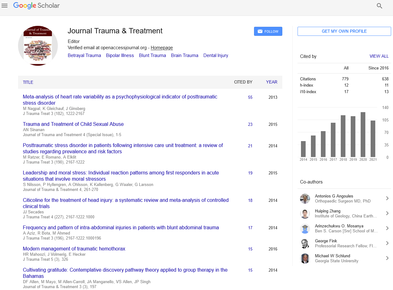
Journal of Trauma & Treatment received 1048 citations as per Google Scholar report