Commentary - (2022) Volume 6, Issue 4
Received: 06-Jul-2022, Manuscript No. jid-22-75172;
Editor assigned: 11-Jul-2022, Pre QC No. P-75172;
Reviewed: 19-Jul-2022, QC No. Q-75172;
Revised: 24-Jul-2022, Manuscript No. R-75172;
Published:
28-Jul-2022
, DOI: 10.37421/2684-4559.2022.6.17 7
Citation: Kahn, Oliver. “The Relationship between Autoimmune
Bullous Dermatoses and Desquamative Gingivitis.” Clin Infect Dis 6 (2022): 177.
Copyright: © 2022 Kahn O. This is an open-access article distributed under the
terms of the Creative Commons Attribution License, which permits unrestricted
use, distribution, and reproduction in any medium, provided the original author
and source are credited.
Desquamative gum disease is an illustrative clinical term described by serious erythema, desquamation, vesicles or bullae prompting disintegrations or ulcers, including the free or joined gingival mucosa. It was first referenced by Books in 1894. In 1932, Prinz laid out it as a spellbinding term for erythema, desquamation, disintegrations and rankles on the minimal and joined gingiva. Following Mc Carthy's theory from 1960, Glickman and Smulowin showed in 1964 that DG is a clinical sign of a few illnesses. The most widely recognized causes are dermatological circumstances, for example, lichen planus (LP) in 70-75% of cases, cicatricial pemphigoid (CP) in 9-14% of cases and pemphigus vulgaris (PV) in 4-13% of cases. Touchiness responses set off by contact allergens in mouthwashes, toothpastes, dental materials or drug may likewise prompt DG. Our story audit centers around the most widely recognized hidden infections appearing with DG, to be specific immune system bullous dermatoses and lichen planus. Immune system bullous infections are a gathering of bleak circumstances with various etiology, pathogenesis and visualization that influence the skin as well as mucous films. The gingival fibromucosa is much of the time the site of beginning of these sicknesses. Hence, the clinical signs might spread to the skin or to other mucous films (conjunctival, nasal, pharyngeal, laryngeal, esophageal or genital). Lichen planus is a persistent provocative mucodermatosis with an immune system component, frequently with oral signs, which can take on, in certain structures, the personality of DG.
As DG is many times the first, on the off chance that by all accounts not the only indication of illness, prompting huge oral uneasiness, patients usually go to the dental specialist for a first conference. It is, in this manner, significant for these experts to know how to analyze the condition, as early satisfactory treatment is fundamental in DG related with immune system illnesses, and a multidisciplinary approach is frequently expected for the suitable administration of these patients [1].
The clinical appearance of DG isn't explicit for a specific illness. Nonetheless, there are a few related clinical discoveries that might highlight an exact finding, which is fundamental, in light of the fact that the sickness course, therapy and forecast are different relying upon the reason [2]. The fundamental contribution and the level of oral mucosal harm are significant variables that add to the overall bleakness and mortality related with immune system problems, as well concerning the general personal satisfaction in these patients.
Different sorts of laser gadgets for instance diodes, helium-neon, bright, to give some examples, can be utilized. These gadgets can be utilized with various boundaries, like result power, season of illumination, portion and conveyed to the oral injury and rehashed for a few meetings. They can be utilized because of their proposed mitigating impacts, relief from discomfort, and sped up recovery of harmed tissues, without showing the antagonistic impacts related with drug admission treatment. This treatment has been upheld because of its mitigating, hostile to bacterial and virucide impacts yet in addition for torment the board and advancing a quicker mending process in the impacted regions, without the unfavorable impacts that medications have [3].
A new meta-examination that broke down 17 investigations has presumed that photobiomodulation decided a general improvement of clinical scores and critical agony decrease of treated regions, like effective corticosteroids, consequently addressing a feasible option [4]. Albeit the fundamental outcomes are promising, these elective treatments ought to be additionally tried and contrasted with the old style treatment to evaluate their adequacy over the long run and for various illness stages.
Another significant perspective that should be considered is the changed personal satisfaction of patients with immune system sicknesses. Studies have featured the connection between the insusceptible framework and the autonomic sensory system through the administrative capability that the last option has on the previous through the parasympathetic and the thoughtful branches. In the event that the autonomic sensory system is impacted, it can create an exacerbated favorable to provocative reaction which can be generally experienced in immune system sicknesses [5]. A few examinations have investigated the effect of immune system sicknesses on personal satisfaction and have reasoned that these patients have an impacted personal satisfaction, expanded pressure, discouragement and nervousness. Infection seriousness can additionally influence personal satisfaction, as there is a positive connection with this record. Besides, a new meta-examination detailed expanded salivary degrees of cortisol, a biomarker for stress and nervousness, in oral lichen planus patients. Taken together, this underscores the requirement for complex administration of these patients, and more consideration ought to be granted to the mental element of treatment.
DG is an obsessive clinical sign of the gingival mucosa that is portrayed by persistent desquamation of the gingival epithelium, erythema, ulceration as well as rankling. This isn't a sickness in itself, yet rather the clinical aggregate of a gathering of illnesses. The most well-known causes are immune system bullous dermatoses, like PV, BP, CP and LP with appearances in the oral mucosa. Some of the time the injuries are completely oral. Thus, early determination of immune system bullous messes is basic for clinicians. A careful clinical assessment, examination of the patient's set of experiences, complete blood count, biochemical tests as well as biopsies are important to figure out a fitting early determination and suggest early treatment with a valuable reaction. Legitimate administration of these patients can decrease secondary effects and lead to a superior guess and personal satisfaction for the patient.
None.
Google Scholar, Crossref, Indexed at
Google Scholar, Crossref, Indexed at
Google Scholar, Crossref, Indexed at
Google Scholar, Crossref, Indexed at
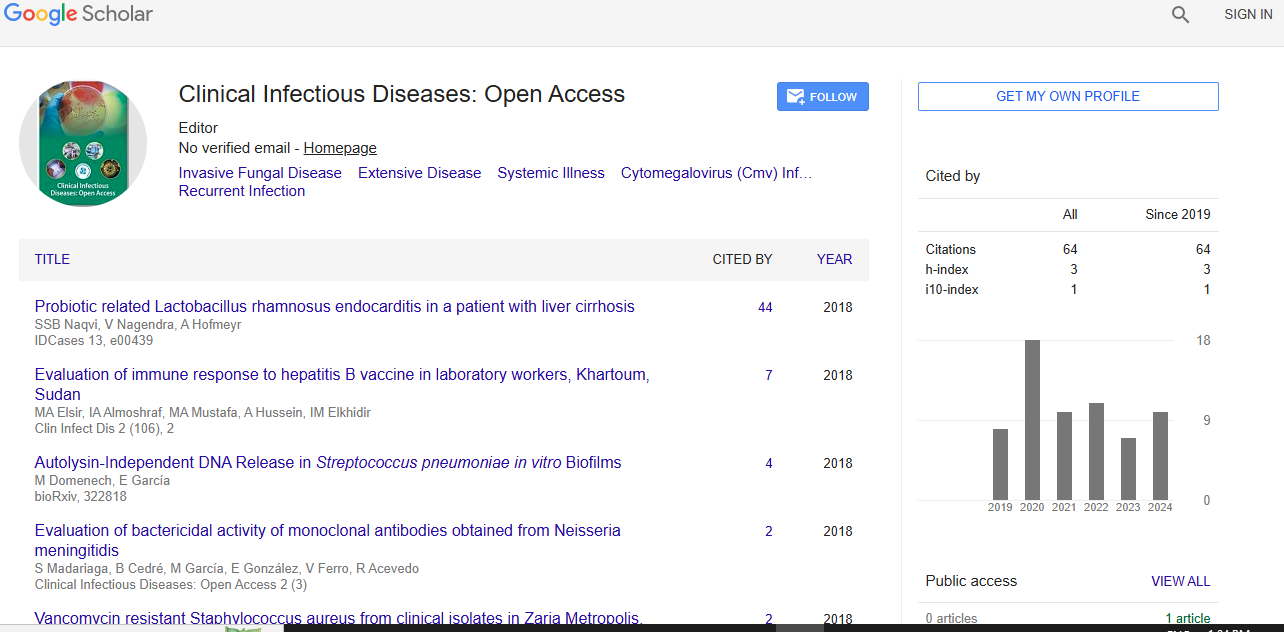
Clinical Infectious Diseases: Open Access received 1149 citations as per Google Scholar report