Research Article - (2026) Volume 12, Issue 2
Received: 08-Apr-2026, Manuscript No. jotr-26-187717;
Editor assigned: 10-Apr-2026, Pre QC No. P-187717;
Reviewed: 25-Apr-2026, QC No. Q-187717;
Revised: 07-May-2026, Manuscript No. R-187717;
Published:
15-May-2026
, DOI: 10.5281/zenodo.20019639
Citation: Jia, Kunpeng , Lexi Huang, Yuanhao Xu and Lingjiao Xiang. “Pan-Cancer Analysis of Molecular Characteristics and Oncogenic Role of PRMT5 in Human Cancers.” J Oncol Transl Res 12 (2026): 341.
Copyright: ©2026 Jia K, et al. This is an open-access article distributed under the terms of the Creative Commons Attribution License, which permits unrestricted use, distribution and reproduction in any medium, provided the original author and source are credited
PRMT5 • Pan-Cancer analysis • Immune infiltration • Prognosis • Proliferation
BRCA: Breast Invasive Carcinoma; ESCA: Esophageal Carcinoma; CCK8: Cell Counting Kit-8; COAD: ColonAdeno Carcinoma; CSEC: Cervical Squamous Cell Carcinoma and Endocervical Adenocarcinoma; HMGB1: High Mobility Group Box 1 Protein; CHOL: Cholangio Carcinoma; KIRC: Kidney Renal Clear Cell Carcinoma; CPTAC: Clinical Proteomic Tumor Analysis Consortium; MSAD: Mucinous Stomach Adenocarcinoma; DFS: Disease-Free Survival; TGCT: Testicular Germ Cell Tumors; DLBC: Diffuse large B-cell lymphoma; PPS: Post Progression Survival; GEO: Gene Expression Omnibus; THCA: Thyroid Carcinoma; GEPIA2: Gene Expression Profiling Interactive Analysis, Version 2; KICH: Kidney Chromophobe; GO: Gene Ontology; OV: Ovarian Serous Cystadenocarcinoma; GTEx: GenoType-Tissue Expression; STAD: Stomach Adenocarcinoma; KEGG: Kyoto Encyclopedia of Genes and Genomes; LUSC: Lung Squamous Cell Carcinoma; LAML: Acute Myeloid Leukemia; READ: Rectum Adenocarcinoma; LGG: Lower Grade Glioma; ACC: Adreno Cortical Carcinoma; LUAD: Lung Adenocarcinoma; GBM: Glio Blastoma Multiforme; OS: Overall Survival; RFS: Relapse-Free Survival; PCNA: Proliferating Cell Nuclear Antigen; FP: First Progression; PRMT5: Protein Arginine Methyl Transferase 5 ; UCEC: Uterine Corpus Endometrial Carcinoma; SAM: S-Adenosine-Methionine; LIHC: Liver Hepatocellular Carcinoma; SKCM: Skin Cutaneous Melanoma; KIRP: Kidney Renal Papillary Cell Carcinoma; TCGA: The Cancer Genome Atlas; HNSC: Head and Neck Squamous Cell Carcinoma; THYM: Thymoma; BLCA: Bladder Urothelial Carcinoma; TIMER2 : Tumor Immune Estimation Resource, Version 2; DSS: Disease-Specific Survival; UCEC: Uterine Corpus Endometrial Carcinoma; PFS: Progress-free Survival
Cancer is a group of diseases characterized by the dysregulation of important pathways that control cellular processes involved in DNA repair, cell survival, proliferation and mortality [1]. With the expanding development of epigenetics, researchers recognize that many biological processes associated with carcinogenesis are epigenetically regulated. Epigenetic alterations such as DNA methylation, histone modification, nucleosome remodeling and RNA-mediated targeting have been demonstrated to drive the onset of cancer [2,3]. Therefore, deciphering aberrant epigenetic mechanisms of tumorigenesis will yield novel insights into anti-cancer strategies.
Arginine methylation mediated by Protein Arginine Methyl Transferase (PRMT) family is a widespread post-translational modification in eukaryotes and plays key roles in many biological processes, such as transcriptional repression Migliori V, et al. [4], RNA splicing Chari A, et al. [5], ribosome biogenesis Ren J, et al. [6], cell death and proliferation [7,8]. Using S-adenosine-methionine as the methyl donor, PRMT transfers methyl groups to arginine side chains and produces S-adenosyl-L-homocysteine and methylarginine [9]. PRMT5, a major methyltransferase of the nine-member PRMT family, has been implicated in oncogenic processes involving both tumor-intrinsic and microenvironmental mechanisms [10]. Although PRMT5 has been examined in several cancer-type-specific studies and database analyses Chung J, et al. [11], its expression, prognosis, immune infiltration and pathway associations have not been systematically integrated across major tumor types. Therefore, a cross-cancer analysis of PRMT5 may help clarify its context-dependent roles and prioritize hypotheses for further functional studies.
The publicly funded TCGA project and the available GEO database contain functional genomics datasets of different tumors [12-14] and thus allow us to conduct pan-cancer analysis. In this study, we aimed to systematically profile PRMT5 across 33 cancer types using TCGA, GTEx and CPTAC data, assess its prognostic and immune-infiltration associations and integrate protein-interaction, KEGG and GO analyses to generate mechanistic hypotheses. We further selected representative cell models to experimentally test whether PRMT5 affects proliferation, migration and stemness-related phenotypes. Together, our pan-cancer analysis delineates a cross-cancer landscape of PRMT5 and provides a framework for future validation.
Data acquisition
PRMT5 gene expression, gene alteration and clinical data of 33 cancer types were obtained from the TCGA repository [15]. Samples lacking PRMT5 expression data, follow-up information, or with concomitant diseases were excluded. Other data were obtained from public databases as described in the corresponding sections.
PRMT5 gene expression analysis
PRMT5 mRNA expression across TCGA tumors and adjacent normal tissues was analyzed using the Tumor Immune Estimation Resource (TIMER 2.0) online tool [16]. For cancer types lacking sufficient normal controls (e.g., lymphoid neoplasm Diffuse Large B-Cell Lymphoma (DLBC), brain Lower Grade Glioma (LGG), Thymoma (THYM)), the expression difference between tumor tissues and corresponding normal tissues in GTEx was determined using the “Expression analysis-Box Plots” module of the GEPIA2 web server Tang Z, et al. [17], under the settings of P-value cutoff = 0.01, log2FC cutoff = 1 and “Match TCGA normal and GTEx data”. In addition, PRMT5 levels across pathological stages (I–IV) were visualized via GEPIA2 violin plots.
To validate findings at the protein level, the UALCAN portal [18] was used to conduct protein expression analysis of the CPTAC [19]. PRMT5 protein expression was compared between primary tumors and normal tissues in the CPTAC dataset. Available datasets from colon cancer, Lung Adeno Carcinoma (LUAD) and Uterine Corpus Endometrial Carcinoma (UCEC) were selected.
Survival prognosis analysis
The Overall Survival (OS) and Disease-Free Survival (DFS) significance map data of PRMT5 across all TCGA tumors were acquired by calculating the log-rank P-value and the HR (hazard ratio; 95% confidence interval) through the “Survival Analysis” module of GEPIA2 [20]. The high-expression and low-expression cohorts were distinguished by cutoff-high (50%) and cutoff-low (50%) values.
Genetic alteration analysis
The cBioPortal for Cancer Genomics, an open platform for interactive exploration of multidimensional cancer genomics data [21,22]. was used to analyze genetic alterations of PRMT5 across all TCGA tumors. The analysis parameters included alteration frequency, mutation type and copy number variation. The mutated site information of PRMT5 was displayed in the schematic diagram of the protein structure of the Three-Dimensional (3D) structure via the “Mutations” module. The overall, disease-specific, progression-free and disease-free survival differences for the TCGA cancer cases with or without PRMT5 mutation were analyzed via the “Comparison” module.
Immune infiltration analysis
The “Immune-Gene module” of TIMER2 web server was employed to analyze the association between PRMT5 expression and immune infiltration across all TCGA cancers. We used the TIMER, EPIC, MCPCOUNTER, CIBERSORT, CIBERSORT-ABS, QUANTISEQ and XCELL algorithms to evaluate immune infiltration. The P-values and partial Correlation (Cor) values were obtained via the purity-adjusted Spearman's rank correlation test. The data were visualized as a heatmap and a scatter plot.
PRMT5-related gene enrichment analysis
The online database STRING website [23] was used to obtain the available experimentally determined PRMT5-binding proteins. The “Similar Gene Detection” module of GEPIA2 was applied to obtain the top 100 PRMT5-related target genes of TCGA database. Then we performed Pearson correlation analysis of PRMT5 and selected genes via the “correlation analysis” module of GEPIA2. Moreover, heatmap data of selected genes was conducted by the “Gene_Corr” module of TIMER2 (Tumor immune estimation resource, version 2) web, which contains the partial correlation (Cor) and P-value in the purity-adjusted Spearman’s rank correlation test.
To further analyze the target genes regulated by PRMT5, we performed an intersection analysis to compare the PRMT5-binding genes and interacted genes via an online Venn diagram tool [24-28]. Moreover, we combined the two sets of data (PRMT5-binding and correlated genes) to perform KEGG pathway analysis. The enriched pathways were finally visualized with “ggplot2” R package. GO enrichment analysis was conducted by the “clusterProfiler” R package. Two-tailed P < 0.05 was considered statistically significant.
Cells and cell culture
Due to the robust endogenous PRMT5 expression and transfection efficiency, Caco-2, HeLa and BGC-823 were selected as representative colorectal, cervical and gastric cancer models for the functional assays, including western blot, proliferation, migration and sphere-formation. All cell lines were routinely maintained in Dulbecco’s modified Eagle’s medium (Hyclone Laboratories Inc., Logan, UT, USA) supplemented with 10% fetal bovine serum (Hyclone Laboratories Inc., Logan, UT, USA) and 1% penicillin-streptomycin (BBI Life Sciences Corporation, Shanghai, China) at 37°C with 5% CO2.
Small interfering RNA transfection
Small interfering RNA (siRNA) was introduced into Caco-2, HeLa and BGC-823 cells at 30-50% confluence using Lipofectamine 3000 transfection reagent (Invitrogen, CA, USA). Commercially available PRMT5 siRNA (5’-CCGCUAUUGCACCUUGGAA-3’) (Gene Pharm, Shanghai, China) and negative control siRNA were used for transfection. Gene silencing was monitored by Western blot assay at 48 h post-transfection.
CCK8 assay
The Caco-2, HeLa and BGC-823 cells were seeded and cultured at a density of 5 × 10^3 /well in 100μL of medium into 96-well microplates (Corning, USA). Then, the cells were transfected with PRMT5 siRNA. Cell proliferation at indicated time points (1 d, 2 d, 3 d and 4 d post-transfection) was evaluated by Cell Counting Kit-8 (CCK8, Beyotime, Shanghai, China) according to the manufacturer's protocols. Optical density values at 460 nm were detected using a microplate reader (Bio-Rad, Hercules, CA, USA).
Quantitative real-time PCR
Total RNA from cells was extracted using Trizol reagent and the RNA was reverse transcribed into cDNA using a reverse transcription reagent kit (RR036A; TaKaRa, Kyoto, Japan), according to the manufacturer's instructions. SYBR® Green qRT-PCR was performed using a Bio-Rad CFX Connect real-time System (CA, USA) to analyze gene expression. The relative levels of each target gene mRNA transcript and control β-actin (housekeeping control) were calculated using the comparative cycle threshold method (2-ΔΔCt) and each experiment was repeated at least 3 times.
Wound healing assay
Caco-2, HeLa and BGC-823 cells treated with PRMT5 siRNA were seeded in 12-well plates. The cell monolayers were wounded by scratching with sterile plastic 200-μl micropipette tips and photographed using phase-contrast microscopy immediately and 24 h after scratching.
Western blot assay
Cells were homogenized in ice-cold lysis buffer RIPA containing protease and phosphatase inhibitor cocktail (Sigma-Aldrich, Shanghai, China). Equal amounts of protein were separated by electrophoresis in SDS-PAGE and then transferred onto nitrocellulose filter membranes (GE whatman, USA). After blocking with 5% nonfat dried milk and then incubated overnight at 4°C with anti-PRMT5 mAb (Cat# ab109451, 1:10,000; Abcam, Shanghai, China), anti-PCNA mAb (Cat# A12427, 1:1000; ABclonal, Wuhan, China), anti-HMGB1 mAb (Cat# A19529, 1:1000; ABclonal, Wuhan, China), anti-p21 mAb (Cat# A19094, 1:1000; ABclonal, Wuhan, China) and anti-β-actin mAb (Cat# AC038, 1:50,000; ABclonal, Wuhan, China). After being washed with Tris-buffered saline with 0.1% Tween (TBST), the membranes were incubated with horseradish peroxidase-conjugated anti-rabbit antibody (Cat# 65-6120, dilution 1:5000; Thermo Fisher, USA) for 2 h at room temperature. The reaction was followed by enhanced chemiluminescence reaction (Cat. WBKLS0500, Millipore, USA) and the blots were quantified using Bio-Rad imaging system (Image Lab 6.0) with β-actin as the internal control.
Tumorsphere-forming assay
A total of 5,000 or 10,000 cells were seeded into 6-well ultralow-attachment plates. For the tumorsphere-formation assay, cells were plated in serum-free DMEM/F12 medium supplemented with B27 and N2 supplement (Invitrogen), human recombinant epidermal growth factor (40ng/mL) and basic fibroblast growth factor (20ng/mL). Media in all dishes were refreshed every 2-3 days. The tumor spheres were photographed after culturing for 7-10 days.
Statistical analysis
For the PRMT5 silencing experiment, quantitative data are expressed as a mean ± standard deviation (s.d.). All data were analyzed using GraphPad Prism 8 software (GraphPad, La Jolla, CA, USA). Statistical analysis of differences was performed by the Student’s t-test between two groups and one‐way analysis of variance followed by Tukey’s test when comparing among multiple groups. Significance was assessed at a level of P < 0.05.
Pan-cancer analysis of PRMT5 expression levels
To evaluate the pan-cancer expression profile of PRMT5, we analyzed its mRNA levels across TCGA cohorts using TIMER2. As shown in Figure 1A, PRMT5 was significantly upregulated in a broad spectrum of malignancies, particularly in BRCA, COAD, ESCA, HNSC, LIHC and LUAD (P <0.001). Similar overexpression was observed in GBM (P <0.01), as well as BLCA and SKCM (P <0.05).
For TCGA cancer types lacking adequate normal controls, we integrated GTEx database samples to supplement the analysis. PRMT5 was markedly elevated in DLBC and THYM, but notably downregulated in LAML (P < 0.05, Figure 1B). No significant expression differences were found in other tumors, including ACC, OV and TGCT (data not shown). To validate these findings at the protein level, analysis of the CPTAC dataset confirmed that PRMT5 protein expression was significantly higher in primary tissues of colon cancer, LUAD and UCEC compared to normal controls (Figure 1C). Finally, we explored the correlation between PRMT5 expression and pathological stages via GEPIA2. PRMT5 levels significantly varied across clinical stages in KICH, KIRC and LIHC (P <0.05, Figure 1D), while no significant stage-associated trends were observed in some other cancer types.

Figure 1. The expression of PRMT5 in different cancers and pathological stages. A). The mRNA expression level of PRMT5 between tumor and normal tissues were compared using the TCGA database, B). The mRNA expression level of PRMT5 in cancers of DLBC, LAML, LGG, and THYM. For the above four cancers of TCGA project without normal tissues, the corresponding normal tissues of the GTEx database were included as controls, C). The protein expression level of PRMT5 between normal tissues and primary tissues of colon cancer, LUAD and UCEC was analyzed based on CPTAC dataset and D). PRMT5 expression levels were analyzed at the main pathological stages (stage I, stage II, stage III and stage IV) of KICH, KIRC and LIHC based on the TCGA dataset. Log 2 (TPM+1) was applied for log-scale. *P< 0.05, **P<0.01, ***P<0.001.
Prognostic significance of PRMT5 expression in human cancers
To evaluate the prognostic significance of PRMT5, we stratified cancer cases from TCGA and GEO databases into high- and low-expression groups. Survival analysis using GEPIA2 revealed that elevated PRMT5 expression was significantly correlated with poorer Overall Survival (OS) in several malignancies, including ACC, LIHC and PAAD (P <0.05, Figure 2A). Similarly, high PRMT5 levels were associated with worse Disease-Free Survival (DFS) in ACC, BLCA and LIHC (Figure 2B). Interestingly, an opposite trend was observed in GBM, KIRC and LGG, where high PRMT5 expression predicted favorable OS and DFS outcomes (P <0.05).

Figure 2. Correlation between PRMT5 expression and survival prognosis of cancers using the TCGA dataset. GEPIA2 was used to determine the correlation of PRMT5 expression with overall survival A). And disease-free survival and B). Prognosis across cancer types. The survival map and Kaplan-Meier curves with positive results are given.
Further validation using the Kaplan-Meier plotter confirmed the diverse prognostic potential of PRMT5 across different cohorts. High PRMT5 expression was linked to inferior Relapse-Free Survival (RFS) in breast cancer and poor OS in lung and liver cancers. Conversely, it indicated a favorable prognosis for gastric cancer (OS and FP) and ovarian cancer (PFS). These findings were largely supported by our meta-analysis, which solidified the prognostic correlation in liver, lung and gastric cancers. Collectively, these data demonstrate that PRMT5 is a context-dependent prognostic biomarker across various human cancers.
Genetic mutation of PRMT5 and its association with survival prognosis in human cancers
To investigate the genetic landscape of PRMT5, we analyzed its mutation status across TCGA cohorts using cBioPortal. As shown in Figure 3A, the highest alteration frequency (>4%) occurred in melanoma, predominantly featuring mutations. Notably, sarcoma and Uterine Carcino Sarcoma (UCS) cases exhibited approximately 2% alteration rates, primarily characterized by gene amplification (Figure 3A). Further mapping of mutation types and sites revealed that missense mutations were the most prevalent, with the R534H/C substitution (Arginine to Histidine/Cysteine) identified in COAD, MSAD and UCEC (Figure 3B). The spatial location of the R534 site was visualized in the 3D protein structure (Figure 3C). Finally, we evaluated the prognostic impact of these alterations although PRMT5 alteration frequency was low (Figure 3D). In UCEC, patients with PRMT5 alterations showed significantly favorable Progression-Free Survival (PFS) (P = 0.0254), although no significant correlations were found for OS, DSS, or DFS. These findings suggest that while PRMT5 mutations are relatively rare, they may possess context-specific prognostic implications.

Figure 3. Mutation feature of PRMT5 in various cancers of TCGA. cBioPortal tool was used to analyze the mutation features of PRMT5 for the TCGA cancers. A). Alteration frequency and mutation types; B). Mutation site, C). Mutation site with the highest alteration frequency (R534) was displayed in the 3D structure of PRMT5 and D). The correlation between mutation status of PRMT5 and OS, DSS, DFS and PFS of UCEC.
Correlation of PRMT5 expression with immune infiltration
Given that the Tumor Micro Environment (TME) plays a critical role in cancer progression and therapy resistance, we investigated whether PRMT5 expression influences the landscape of tumor-infiltrating immune and stromal cells. Specifically, we focused on CD8+ T cells, the primary effectors of anti-tumor immunity and Cancer-Associated Fibroblasts (CAFs), which are known to modulate immune exclusion and promote tumor malignancy. Therefore, we utilized multiple deconvolution algorithms, including TIMER, EPIC, MCP-COUNTER, CIBERSORT, QUANTISEQ and XCELL, to evaluate the correlation between PRMT5 expression and the infiltration levels of CD8+ T cells and cancer-associated fibroblasts.
Consistent across most algorithms, PRMT5 expression was negatively correlated with CD8+ T-cell infiltration in CESC and SKCM (Figure 4), suggesting a potential role in immune evasion. Furthermore, we observed a significant positive correlation between PRMT5 and CAFs across various TCGA tumors, such as ACC, CESC, CHOL, LIHC and PAAD (Figure 5A). Scatterplot analysis further confirmed these associations; for example, PRMT5 levels in ACC were positively linked to CAF infiltration (Cor = 0.349, P = 2.48e-03) according to the TIDE algorithm (Figure 5B). Interestingly, a negative correlation with CAFs was noted in LGG. These findings indicate that PRMT5 may modulate the TME by influencing immune and stromal cell recruitment.

Figure 4. Correlation analysis between PRMT5 expression and infiltration of CD8+ T cells.

Figure 5. Correlation analysis between PRMT5 expression and infiltration of cancer-associated fibroblasts.
Enrichment analysis of PRMT5-related partners
To further explore the molecular basis by which PRMT5 modulates tumor progression and the immune microenvironment, we constructed a Protein-Protein Interaction (PPI) network using the STRING tool. Based on experimental evidence, 44 PRMT5-binding proteins were identified (Figure 6A). Simultaneously, we identified the top 100 genes significantly correlated with PRMT5 expression via GEPIA2. As shown in Figure 6B, PRMT5 expression was strongly and positively associated with genes such as Ty 16 homolog (R = 0.69), 5 proteasome (R = 0.64), Neuroguidin (R = 0.66), nucleolin (NCL) (R = 0.48), Eukaryotic Translation Initiation Factor 2 subunit 1 (EIF2S1) (R = 0.52) and apyrimidinic endonuclease 1 (R = 0.62). Heatmap analysis further confirmed these positive correlations across various cancer types (Figure 6C). Notably, intersection analysis via a Venn diagram revealed that EIF2S1 and NCL were both physically bound to and expression-correlated with PRMT5 (Figure 6D), which were well-recognized regulators involved in RNA processing, translation initiation and genomic stability.
To better characterize the biological roles of these 142 PRMT5-associated genes (binding and correlated), KEGG and GO enrichment analyses were performed. The most significantly enriched KEGG pathways included "systemic lupus erythematosus", "viral carcinogenesis" and the "spliceosome" (Figure 6E). Furthermore, GO annotation indicated that these genes are primarily involved in DNA/RNA metabolism and stress response, including activities such as catalytic activity on DNA/RNA, ATPase activity and Hsp90 protein binding (Figure 6F). These results suggest that PRMT5 may exert its oncogenic effects by modulating RNA processing and genomic stability.

Figure 6. PRMT5-related gene enrichment analysis. A). The available experimentally determined PRMT5-binding proteins, B). The expression correlation of PRMT5 and selected target genes from the top 100 PRMT5-correlated genes in TCGA projects, C). The corresponding heatmap data of the correlation analysis in detailed cancer types, D). An intersection analysis of PRMT5-correlated and PRMT5-binding genes. KEGG pathway, E). and GO analysis and F) based on the PRMT5-correlated and binding genes.
PRMT5 depletion decreases the stemness, proliferative and migratory capacities of carcinoma cells
Considering that PRMT5-associated genes are predominantly enriched in transcription coregulation and RNA metabolism, two fundamental processes governing cellular plasticity, we hypothesized that PRMT5 may serve as a critical regulator of cancer cell stemness and aggressive phenotypes to support cancer cell proliferation and related phenotypes.
To test this, we performed loss-of-function assays in Caco-2, HeLa and BGC-823 cells. PRMT5 silencing significantly impaired cell proliferation (Figure 7A) and led to a marked downregulation of stemness-associated markers, including ALDH1A1, CD44, OCT4 and SOX9 (Figure 7B). Consistently, the tumorsphere-forming efficiency, a hallmark of self-renewal, was significantly reduced in the siPRMT5 group compared to the control (Figure 7C).
Beyond stemness, we assessed the impact of PRMT5 on migratory capacity. Wound healing assays demonstrated that PRMT5-knockdown cells exhibited a reduced capacity to close the initial scratch area (Figure 7D). Mechanistically, the observed suppression of proliferation and migration was accompanied by decreased protein levels of PCNA, p21 and HMGB1 (Figure 7E), which are key regulators of the cell cycle and motility. Collectively, these data provide experimental evidence that PRMT5 is essential for maintaining the stemness and metastatic potential of tumor cells, validating our initial bioinformatic predictions.

Figure 7. Knockdown of PRMT5 inhibited stemness and migration of tumor cells. Tumor cells were transfected with PRMT5 siRNA (siPRMT5) or scrambled siRNA (siScr) as described in Materials and Methods. A). The proliferation of Caco-2, HeLa, and BGC-823 cells was examined by CCK-8 assay, B). qRT-PCR of ALDH1A1, CD44, OCT4, and SOX9 relative gene expression normalized to β-actin in Caco-2, HeLa, and BGC-823 cells C). Tumorsphere-forming assay micrograph of Caco-2, HeLa, and BGC-823 cells after culturing for 10 days, scale bar 100 μm, D). The migration of Caco-2, HeLa, and BGC-823 cells was examined by healing assay. Scale bar, 100 μm and E). Western blot and quantitative analyses of PCNA, p21 and HMGB1 in control and PRMT5-silenced Caco-2, HeLa, and BGC-823 cells. Data were shown as mean ± s.d. *P < 0.05, **P < 0.01, ***P < 0.001, ****P < 0.0001.
In this study, we systematically characterized the dysregulation and oncogenic potential of PRMT5 across 33 cancer types, providing a comprehensive cross-cancer framework illustrating the clinical and biological significance of PRMT5. Our findings demonstrate that while PRMT5 is predominantly upregulated across human malignancies, its impact on patient prognosis and the Tumor Micro Environment (TME) is highly context-dependent.
Consistent with previous reports in colorectal and hepatic cancers [29-35], our analysis confirms the widespread overexpression of PRMT5 in the majority of TCGA cohorts. PRMT5 is traditionally viewed as an oncogene associated with poor survival [36-39]. However, we discovered a prognostic paradox that elevated PRMT5 correlates with prolonged survival in GBM, KIRC and LGG, contrasting sharply with its role in ACC, LIHC and PAAD. This discrepancy can be partly interpreted that PRMT5 regulates the expression of genes implicated in both tumor promotion and suppression [40]. For another, previous evidence suggests that PRMT5’s functional output is dictated by its subcellular localization [41]. Nuclear PRMT5 has been associated with growth inhibition and favorable prognosis in breast and prostate cancers, whereas its cytoplasmic counterpart drives oncogenesis [42-44]. Thus, the protective role observed in gliomas and renal cancers likely reflects tissue-specific differences in PRMT5 sequestration or substrate availability. By highlighting these divergent prognostic values within a unified analytical framework, we underscore the necessity of considering cancer-specific backgrounds in the development of PRMT5-targeted therapies.
A pivotal finding of our study is the potential role of PRMT5 in remodeling the Tumor Micro Environment (TME). We observed a consistent negative correlation between PRMT5 expression and CD8+ T-cell infiltration in CESC and SKCM, suggesting a contribution to immune evasion. This aligns with recent evidence that pharmacological inhibition of PRMT5 by DS-437 can "re-awaken" the immune response by increasing T-cell recruitment, thus reducing tumor growth in breast cancer [45,46]. Furthermore, we identified a positive association between PRMT5 and Cancer-Associated Fibroblasts (CAFs) across multiple tumors. CAFs are major components of the tumor stroma that promote malignancy by constructing physical barriers against immune cell entry and secreting pro-tumorigenic factors [47,48]. The dual observation of decreased CD8+ T cells and increased CAF infiltration suggests that PRMT5 may facilitate the formation of "cold tumors", an environment characterized by immune exclusion. Deciphering the PRMT5-CAF axis will be crucial for developing combinatorial strategies that pair PRMT5 inhibitors with current immunotherapies.
Mechanistically, our protein-protein interaction and enrichment analyses identified PRMT5 as a central hub for DNA/RNA metabolism and spliceosome assembly. Notably, we found that PRMT5 physically interacts and co-expresses with NCL and EIF2S1, which are well-recognized regulators of RNA processing and translation initiation. Increasing evidence suggests that DNA and RNA modification are mis-regulated in human cancers and are ideal targets of cancer therapy [49,50]. Interestingly, a flourish of recent reports have solidified PRMT5 as a major regulator of epigenetic-mediated gene expression Migliori V, et al. [51], mRNA splicing Koh CM, et al. [52] and the DNA damage response Hamard PJ, et al. [53] in the setting of tumors. Our observation that PRMT5 knockdown reduced stemness markers and tumorsphere formation is consistent with these RNA-processing and transcription-related annotations. Additionally, Fong et al. (2019) demonstrated that inhibiting symmetric dimethylation of arginine mediated by PRMT5 reduces RNA splicing catalysis and results in preferential killing of leukemias [54]. Along with DNA/RNA modification, excessive proliferation and migration of cancer cells are involved in tumorigenesis and metastasis [55]. Herein, we demonstrated that PRMT5 knockdown repressed the proliferative and migrative capacities of gastric cancer cells by modulating critical regulators PCNA, p21 and HMGB1. Consistently, several studies have also linked PRMT5 to liver carcinogenesis by promoting cell growth, migration and invasion [56,57]. Thus, the above findings together suggested that PRMT5 is a key epigenetic regulator of cancer cell biology and may contribute to cancer development.
In conclusion, our systematic pan-cancer analysis demonstrates that PRMT5 is predominantly upregulated across human malignancies. Its dysregulation is significantly associated with clinical prognosis, CD8+ T-cell and cancer-associated fibroblast infiltration and the genetic mutational landscape in a cancer-type-specific manner. Furthermore, mechanistic insights suggest that PRMT5 contributes to tumor progression by modulating DNA/RNA metabolism, the spliceosome and stress-response pathways, thereby maintaining cancer cell stemness and aggressive phenotypes. Our study highlights the importance of considering the specific molecular and immune context when developing PRMT5-targeted strategies for personalized cancer therapy.
Originality Statement
The authors declare that this manuscript is original, has not been published or submitted elsewhere in whole or in part and is not under consideration for publication by any other publisher.
The authors declare that they have no competing interests
Lingjiao Xiang: Writing–review & editing, Writing–original draft, Visualization, Validation, Methodology, Investigation, Formal analysis, Data curation, Conceptualization, Software, Resources.
Kunpeng Jia: Writing–review & editing, Writing–original draft, Visualization, Validation, Methodology, Investigation, Formal analysis, Data curation, Conceptualization.
Lexi Huang: Writing–review & editing, Methodology, Investigation, Formal analysis, Data curation, Conceptualization. Yuanhao Xu: Writing–review & editing.
Google Scholar, Cross Ref, Indexed at
Google Scholar, Cross Ref, Indexed at
Google Scholar, Cross Ref, Indexed at
Google Scholar, Cross Ref, Indexed at
Google Scholar, Cross Ref, Indexed at
Google Scholar, Cross Ref, Indexed at
Google Scholar, Cross Ref, Indexed at
Google Scholar, Cross Ref, Indexed at
Google Scholar, Cross Ref, Indexed at
Google Scholar, Cross Ref, Indexed at
Google Scholar, Cross Ref, Indexed at
Google Scholar, Cross Ref, Indexed at
Google Scholar, Cross Ref, Indexed at
Google Scholar, Cross Ref, Indexed at
Google Scholar, Cross Ref, Indexed at
Google Scholar, Cross Ref, Indexed at
Google Scholar, Cross Ref, Indexed at
Google Scholar, Cross Ref, Indexed at
Google Scholar, Cross Ref, Indexed at
Google Scholar, Cross Ref, Indexed at
Google Scholar, Cross Ref, Indexed at
Google Scholar, Cross Ref, Indexed at
Google Scholar, Cross Ref, Indexed at
Google Scholar, Cross Ref, Indexed at
Google Scholar, Cross Ref, Indexed at
Google Scholar, Cross Ref, Indexed at
Google Scholar, Cross Ref, Indexed at
Google Scholar, Cross Ref, Indexed at
Google Scholar, Cross Ref, Indexed at
Google Scholar, Cross Ref, Indexed at
Google Scholar, Cross Ref, Indexed at
Google Scholar, Cross Ref, Indexed at
Google Scholar, Cross Ref, Indexed at
Google Scholar, Cross Ref, Indexed at
Google Scholar, Cross Ref, Indexed at
Google Scholar, Cross Ref, Indexed at
Google Scholar, Cross Ref, Indexed at
Google Scholar, Cross Ref, Indexed at
Google Scholar, Cross Ref, Indexed at
Google Scholar, Cross Ref, Indexed at
Google Scholar, Cross Ref, Indexed at
Google Scholar, Cross Ref, Indexed at
Google Scholar, Cross Ref, Indexed at
Google Scholar, Cross Ref, Indexed at
Google Scholar, Cross Ref, Indexed at
Google Scholar, Cross Ref, Indexed at
Google Scholar, Cross Ref, Indexed at
Google Scholar, Cross Ref, Indexed at
Google Scholar, Cross Ref, Indexed at
Google Scholar, Cross Ref, Indexed at
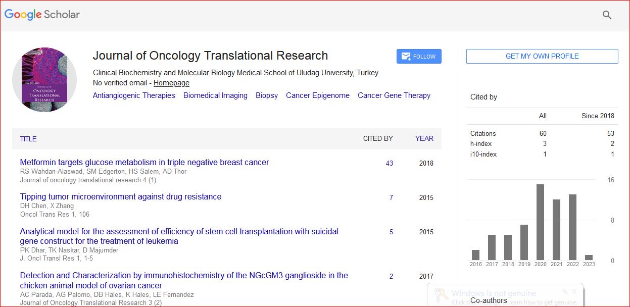
Journal of Oncology Translational Research received 93 citations as per Google Scholar report