Editorial - (2022) Volume 6, Issue 1
Received: 03-Jan-2022, Manuscript No. Jchd-22-57494;
Editor assigned: 05-Jan-2022, Pre QC No. P-57494;
Reviewed: 08-Jan-2022, QC No. Q-57494;
Revised: 13-Jan-2022, Manuscript No. R-57494;
Published:
18-Jan-2022
, DOI: 10.37421/jchd.2022.6.133
Citation: Marcela, Lina. “Editorial Note on Ventricular Septal Defect.” J Coron Heart Dis 6 (2022):133. DOI: 10.37421/jchd.2022.6.133.
Copyright: © 2022 Marcela L. This is an open-access article distributed under the terms of the Creative Commons Attribution License, which permits unrestricted use, distribution, and reproduction in any medium, provided the original author and source are credited.
A ventricular septal defect (VSD), also known as a hole in the heart, is a common birth defect (congenital). The hole (defect) occurs in the wall (septum) that separates the lower chambers (ventricles) of the heart, allowing blood to flow from the left to the right side of the heart. The oxygen-rich blood is then pumped back to the lungs rather than out to the body, making the heart work harder. A minor ventricular septal defect may not cause any issues, and many minor VSDs close on their own. To avoid complications, medium or larger VSDs may require surgical repair early in life. Signs and symptoms of serious heart defects frequently appear during a child's first few days, weeks, or months of life. Poor eating, failure to thrive, and other symptoms of a ventricular septal defect (VSD) in a baby can occur. Breathing too quickly or too slowly, Simple exhaustion.
Congenital heart defects are caused by problems early in the development of the heart, but there is often no clear cause. Environmental and genetic factors may both play a role. VSDs can occur on their own or in conjunction with other congenital heart defects. A ventricular septal defect occurs during foetal development when the muscular wall separating the heart into left and right sides (septum) fails to form completely between the lower chambers of the heart (ventricles). Normally, the right side of the heart sends oxygen-rich blood to the lungs, while the left side sends oxygen-depleted blood to the rest of the body.
A VSD allows oxygenated and deoxygenated blood to mix, resulting in increased blood pressure and blood flow in the lung arteries. VSDs come in a variety of sizes and can be found in a variety of locations in the wall between the ventricles. One or more VSDs may exist. A VSD can also develop later in life, usually as a result of a heart attack or as a complication from certain heart procedures. Ventricular septal defects can run in families and can coexist with other genetic disorders such as Down syndrome.
If you already have a child with a heart defect, a genetic counsellor can talk with you about the possibility of your next child having one as well. A minor ventricular septal defect may never cause any complications. Medium and large defects can result in a variety of disabilities ranging from mild to life threatening. Many complications can be avoided with treatment. Failure of the heart, when a heart has a medium or large VSD, the heart works harder and the lungs receive an excessive amount of blood. Heart failure can develop if not treated. Hypertension of the lungs, Because of the VSD, increased blood flow to the lungs causes high blood pressure in the lung arteries (pulmonary hypertension), which can permanently damage them [1-5].
This complication may result in blood flow through the hole being reversed (Eisenmenger syndrome) Endocarditis.This type of heart infection is a rare complication. Other heart issues these include irregular heartbeats and valve issues. In most cases, there is nothing you can do to avoid having a baby with a ventricular septal defect. However, it is critical to take every precaution to ensure a healthy pregnancy. Get prenatal care as soon as you find out you're pregnant. Before becoming pregnant, discuss your health with your doctor and any lifestyle changes that your doctor may recommend for a healthy pregnancy. Also, make sure to discuss any medications you're taking with your doctor.
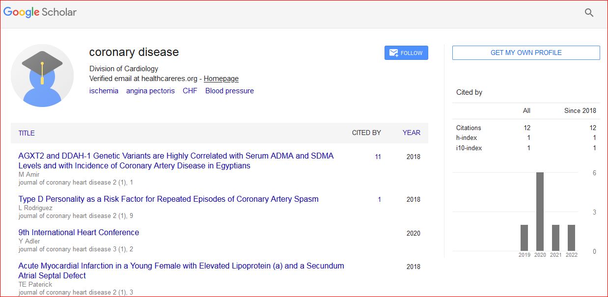
Journal of Coronary Heart Diseases received 15 citations as per Google Scholar report