Opinion - (2022) Volume 13, Issue 1
Received: 19-Jan-2022, Manuscript No. jbsbe-22- 56039;
Editor assigned: 21-Jan-2022, Pre QC No. P-56039;
Reviewed: 27-Jan-2022, QC No. Q-56039;
Revised: 03-Feb-2022, Manuscript No. R-56039;
Published:
08-Feb-2022
, DOI: 10.4172/2155-6210.22.13.312
Citation: Lin, Yuehe. "DNA Detection by using a Bio-microfluidic Interface." J Biosens Bioelectron 13 (2022): 312.
Copyright: © 2022 Lin Y. This is an open-access article distributed under the terms of the Creative Commons Attribution License, which permits unrestricted use, distribution, and reproduction in any medium, provided the original author and source are credited.
Microfluidics has exploded in popularity in recent years, enabling costeffective lab-on-a-chip platforms for drug discovery, cell and molecular interactions research, and, most importantly, molecular diagnostics. Microfluidic devices enable the use of tiny quantities of reagents and samples in high-throughput portable systems with the potential for automation and highlevel integration, as well as cost savings. These devices may also be built for multi-parallel operation, which increases the system's reliability by allowing numerous control assays to be run simultaneously with multiple samples.
Many microfluidic devices for DNA analysis have recently been created, enabling for miniaturised DNA amplification, detection of picograms of DNA, rapid DNA hybridization, and even integrated DNA analysis, which includes sample pretreatment, DNA amplification, and detection. However, due to the high cost of production and/or modification, a comprehensive lab on chip device for DNA analysis has yet to be marketed. Nanodiagnostics, or the use of nanotechnology platforms for nanosensing applications based on gold nanoparticles' extraordinary optical capabilities, has revolutionised the molecular area of examination. For DNA detection, the use of thiol-ssDNA functionalised gold nanoparticles paved the path for simple yet sensitive and specific molecular diagnostic techniques.
Among these, a detection scheme based on the differential colorimetric behaviour of Au-nanoprobes after salt induced aggregation mediated by the presence of a complementary target sequence has been widely investigated - the presence of a complementary target prevents aggregation and the solution retains its original red colour; however, the absence of this specific sequence causes aggregation and the solution turns blue. This colorimetric detection system was used to successfully identify Mycobacterium tuberculosis, the major etiological agent of human TB, which affects over 8.3 million people globally.
This approach has been improved to allow for the detection and characterization of drug resistance mutations as well as the identification of M. tuberculosis Complex members without the requirement for target amplification. Green and red light sources, as well as tiny p-i-n silicon or TiO2- based ink-jet printed photodetectors, have been successfully combined into an easy-to-operate, affordable, and dependable optoelectronic platform, and more recently with paper-based microfluidics. As the first step toward an integrated lab-on-chip device for point-of-care usage, we present the creation of a biomicrofluidic platform for colorimetric DNA analysis based on Au-nanoprobes.
Colorimetric measurements in microfluidic systems are difficult to accomplish because a reduction in the optical path length inside the microchannel system might reduce sensitivity, especially when the channel depth is utilised. To do so, we used optical fibres to carry light from the source to the microchannel and then to the photodetector, extending the OPL by detecting along a microchannel.
The insertion grooves established in the microfluidic chip self-align the optical 4 fibres with each other and with the detecting channel. To minimise cross contamination, the microfluidic chip with optical fibres may be discarded after each test, whereas the green and red light emitting diodes, detector, and electrical setup are permanent components of the biosensor system. The chip is made of polydimethylsiloxane, a silicon rubber that was chosen for its low cost, biocompatibility, strong optical qualities, and ability to replicate micrometer-scale features with great fidelity via replica moulding.
For the suggested design, PDMS must be patterned using a mould that allows for the creation of high-aspect-ratio features such as high grooves for fibre insertion and smooth, vertical side walls to decrease optical losses in the system. Although SU-8, an epoxy-based negative photoresist, meets these requirements, SU-8 moulds experience delamination at the photoresistsubstrate interface after a few PDMS copies, necessitating a repeat of the time-consuming and costly SU-8 photolithography.
Aside from that, SU-8 is extremely sensitive to processing factors, making it difficult to make two identical SU8 samples. It is preferable to create numerous PDMS chips from the same mould in order to obtain excellent repeatability. To tackle this, we employed an intermediate epoxy mould that had previously been used to process 20 m tall features to process our 125 m features, and we experimentally validated that this technique could be used to produce taller structures [1-5].
Following the construction, characterisation, and optimization of this microfluidic technology, it was employed to detect a particular DNA target sequence capable of unambiguous identification of MTBC members. We were able to achieve a specific identification of DNA from M. tuberculosis using just 3 l of solution utilising the suggested microfluidic technology, which represents a 20-fold reduction in volume when compared to the current state of the art.
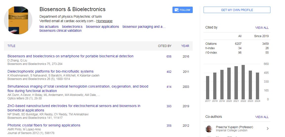
Biosensors & Bioelectronics received 6207 citations as per Google Scholar report