Opinion - (2022) Volume 6, Issue 3
Received: 01-Jun-2022, Manuscript No. jcao-22-71734;
Editor assigned: 03-Jun-2022, Pre QC No. P-71734;
Reviewed: 08-Jun-2022, QC No. Q-71734;
Revised: 13-Jun-2022, Manuscript No. R-71734;
Published:
18-Jun-2022
, DOI: 10.37421/2684-6004.2022.6.140
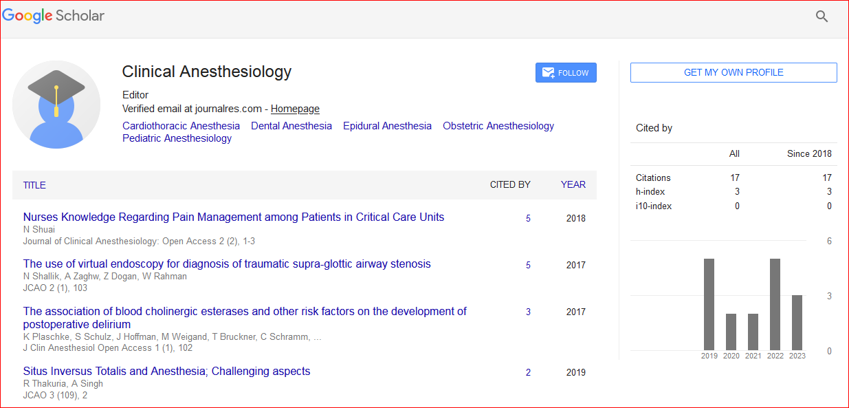
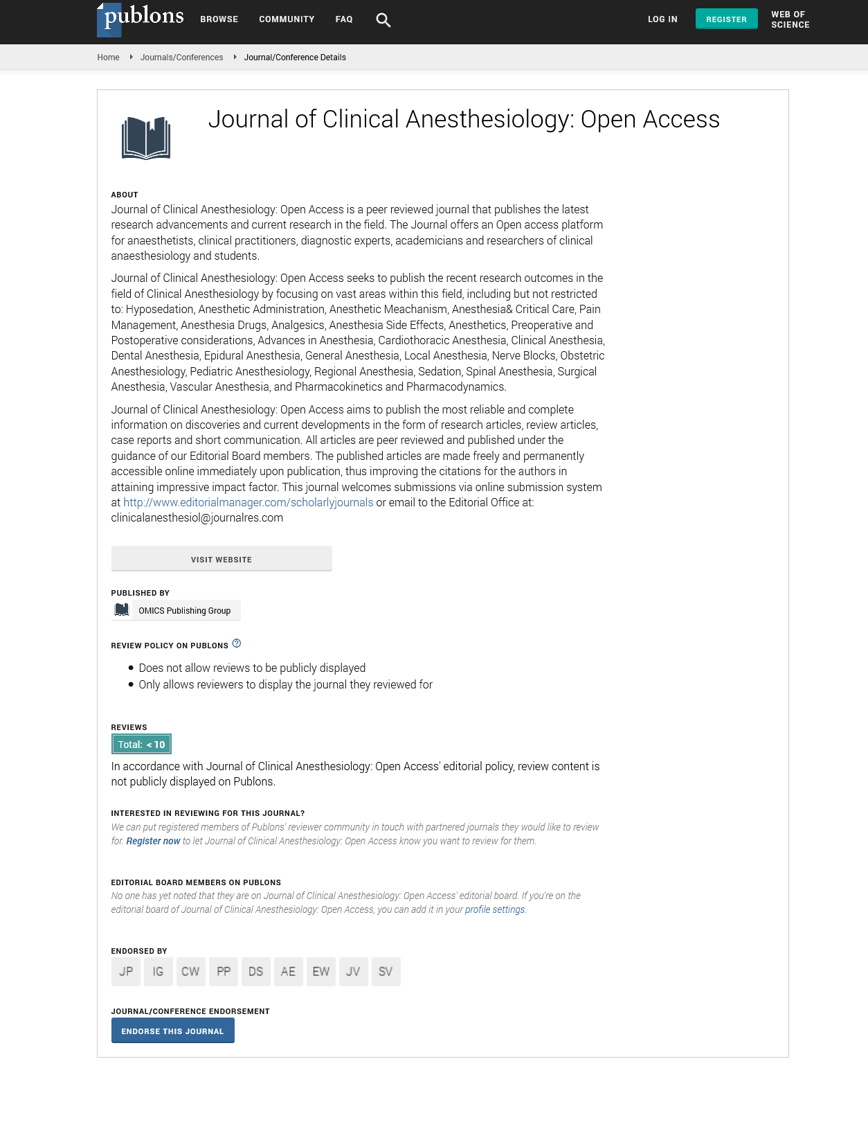
Citation: Guasch, Emilia. “Consideration of Hypoxemia during Anaesthesia and Surgical Treatment.” J Clin Anesthesiol 6 (2022): 140.
Copyright: © 2022 Guasch E. This is an open-access article distributed under the terms of the Creative Commons Attribution License, which permits unrestricted use, distribution, and reproduction in any medium, provided the original author and source are credited.
Because oxygen is essential to life, humans can only exist without it for a short period of time. A balance between oxygen demand and delivery is necessary for the body to maintain homeostasis. The two primary organ systems in charge of supplying the body with oxygen and preserving homeostasis are the respiratory and cardiovascular systems. Any abnormality in either of these two processes would lead to hypoxemia and its detrimental effects. A ventilation/perfusion mismatch is the most frequent underlying cause of hypoxemia, while there are other potential causes as well. The focus of the current review will be on how hypoxemia in humans is defined, caused by what, how it's treated, etc. In contrast to hypoxia, which is marked by a decrease in tissue oxygenation, hypoxemia is characterised by a fall in the partial pressure of oxygen in the blood. It might be brought on by either insufficient oxygen delivery or insufficient oxygen uptake by the tissues. Hypoxemia and hypoxia don't always go along. Patients may have hypoxemia without hypoxia if their haemoglobin level and cardiac output increase in a compensatory manner (CO). It is also possible to have hypoxia without hypoxemia. Cells with cyanide poisoning cannot utilise oxygen despite normal blood and tissue oxygen levels. It is well recognised that flexible bronchoscopy can result in oxygen desaturation, which is more common when bronchoalveolar lavage is performed. When done in a safe environment, flexible bronchoscopy is a treatment with few severe side effects. Flexible bronchoscopy can result in substantial hypoxemia despite the use of additional oxygen. In an emergency, hypoxemia can cause symptoms including breathing pain. Examples include having trouble breathing, inhaling more quickly, using your chest and abdomen to breathe, and pursing your lips. Depending on the situation, chronic hypoxemia may be compensated or uncompensated. The compensation may initially mask symptoms, but additional illness or stress, such as a rise in oxygen demand, may eventually make the hypoxemia more apparent.
Blood vessels that supply less well-ventilated portions of the lungs may selectively contract in a compensated state to reroute blood to betterventilated parts of the lungs. This procedure, however, can result in pulmonary hypertension, overloading the right ventricle of the heart, cor pulmonale, and right sided heart failure if the lungs are not generally ventilated adequately. The risk of polycythemia is another. In addition to stunted growth, poor sleep quality, and frequent sleep arousals, hypoxia can also affect a person's neurological and motor development. Cyanosis, digital clubbing, and symptoms connected to the source of the hypoxemia, such as cough and hemoptysis, are some other signs of hypoxia. When the partial pressure of oxygen in blood is less than 60 mmHg, the steep section of the oxygen-hemoglobin dissociation curve begins, where a small decrease in the partial pressure of oxygen results in a substantial decrease in the oxygen content of the blood. Severe hypoxia can lead to respiratory failure. Hypoxemia is characterised as low blood oxygen levels. Any factor that alters the rate or quantity of air entering the lungs (ventilation) or the transfer of air from the lungs to the blood might result in hypoxemia. Numerous variables, such as cardiovascular disorders like shunts and respiratory problems, can contribute to hypoxemia. The five etiologies that cause hypoxemia are hypoventilation, ventilation/perfusion mismatch, rightto- left shunt, diffusion impairment, and low partial pressure of oxygen. Low oxygen partial pressure and hypoventilation are related to a normal alveolararterial gradient, but the other categories are related to an elevated A-a gradient. If alveolar ventilation is insufficient, there won't be enough oxygen transferred to the alveoli for the body to use. Hypoxemia can happen even if the lungs are healthy due to an issue with the brainstem's ventilation control or the body's inability to breathe properly.
Centers in the medulla that regulate respiration have an impact on the rate and depth of each breath. The central nervous system's peripheral and central chemoreceptors, as well as the carotid and aortic bodies, respectively, influence how much carbon dioxide present in the blood. Hypoxia happens when the breathing centre isn't functioning properly or when the signal isn't correct. The medullary respiratory centres, which generate rhythmic impulses and transmit them to the diaphragm, the breathing muscle, via the phrenic nerve, can be harmed by strokes, seizures, and cervical neck fractures. A reduction in respiratory drive can also result from metabolic alkalosis, which is a deficiency in blood carbon dioxide. Obstructive sleep apnea includes central sleep apnea. During sleep, the brain's respiratory centres may stop functioning, leading to prolonged episodes of apnea with potentially fatal consequences. followed by a period of holding your breath after hyperventilating. In an effort to reduce the amount of carbon dioxide in their lungs, some swimmers try hyperventilating. The urge to breathe in is lessened as a result. However, it does imply that hypoxemia may develop if declining blood oxygen levels are not noticed. Numerous factors, including V/Q mismatch, right-to-left shunt, diffusion impairment, hypoventilation, and low inspired PO2, can lead to hypoxemia. The conditions that specifically determine the partial pressure of oxygen and partial pressure of carbon dioxide in gas exchange units of the lung in ADP ribosylation factors secondary to chronic obstructive pulmonary disease exacerbations and acute lung injury are the ventilation per perfusion ratio and the composition of inspired gas and mixed venous blood. Acute renal failure secondary to chronic obstructive pulmonary disease exacerbations and the acute respiratory distress syndrome, while the ventilation per perfusion ratio and the composition of inspired gas and mixed venous blood, are the conditions that specifically determine the partial pressure of oxygen and partial pressure of carbon dioxide in gas exchange units of the lung.
First-line treatment for hypoxemic acute respiratory failure is oxygen therapy (ARF). An alternative to traditional oxygen therapy is high-flow nasal oxygen therapy (HFNO). With HFNO, patients get humidified, titrated oxygen therapy that meets or exceeds their inspiratory demand. Intensive Care Units (ICUs) are increasingly using HFNO due to mounting data from multiple research demonstrating its efficacy. It is still unclear how HFNO works and what physiological consequences it has. Potential mechanisms include pharyngeal dead space washout, decreased airway resistance, production of a positive end-expiratory pressure, and improved oxygen delivery. According to recent research, HFNO is helpful in most patients with hypoxaemic ARF of various etiologies in terms of increasing oxygenation. Despite the possibility that HFNO could be helpful in the management of hypoxaemia, more extensive cohort studies are required to define the indications, contraindications, and causes of HFNO failure. In the treatment of post-extubation ARF, HFNO might also be helpful in lowering the requirement for tracheal intubation. Additionally, it has been suggested that HFNO can prevent oxygen desaturation by extending apnoeic oxygenation during intubation in both operating rooms and intensive care units. The practise of anaesthesia depends critically on respiratory function. The proper execution of daily procedures for the induction and maintenance of general anaesthesia, the provision of mechanical ventilation, the discontinuation of mechanical and pharmacologic support, and the return to the preoperative state is aided by knowledge of the fundamental physiologic principles of respiration. The current publication offers a review of traditional physiology and places an emphasis on elements crucial to anesthesiologists.
Gas exchange and respiratory mechanics are the two primary divisions of the content, and in each of these divisions, the physiology is presented as the foundation for abnormal situations. Reviewing the routes taken by oxygen from the air to the arteries and carbon dioxide in the opposite direction, we can determine what causes hypoxemia and hypercarbia based on these very pathways. We outline the normal ventilation-determining activities of pressure, flow, and volume, and we discuss the associated aberrations in terms of variations in resistance and compliance. An interventional pulmonologist must be proficient in rigid bronchoscopy as it is a vital tool for treating airway problems. It has remained a crucial procedure for the treatment of central airway obstruction, foreign body aspiration, and severe hemoptysis since its invention in the late 19th century. regions covered The history, indications, contraindications, technique, and complications of rigid bronchoscopy will all be covered in this article. A brief discussion of anaesthetic and ventilation techniques will also be included, followed by our outlook on the future of rigid bronchoscopy. Expert analysis: Although rigid bronchoscopy usage fell after the development of flexible bronchoscopy in the 1960s, it has seen a return during the past two decades. We believe it will continue to be a crucial tool utilised by interventional pulmonologists for many years to come. We propose that interventional pulmonologists need to receive training and gain proficiency in this approach [1-6].
None.
Google Scholar, Crossref, Indexed at
Google Scholar, Crossref, Indexed at
Google Scholar, Crossref, Indexed at
Journal of Clinical Anesthesiology: Open Access received 31 citations as per Google Scholar report