Perspective - (2022) Volume 12, Issue 10
Received: 01-Oct-2022, Manuscript No. jbpbt-23-85927;
Editor assigned: 03-Oct-2022, Pre QC No. P-85927;
Reviewed: 15-Oct-2022, QC No. Q-85927;
Revised: 20-Oct-2022, Manuscript No. R- 85927;
Published:
27-Oct-2022
, DOI: 10.37421/2155-9821.2022.12.544
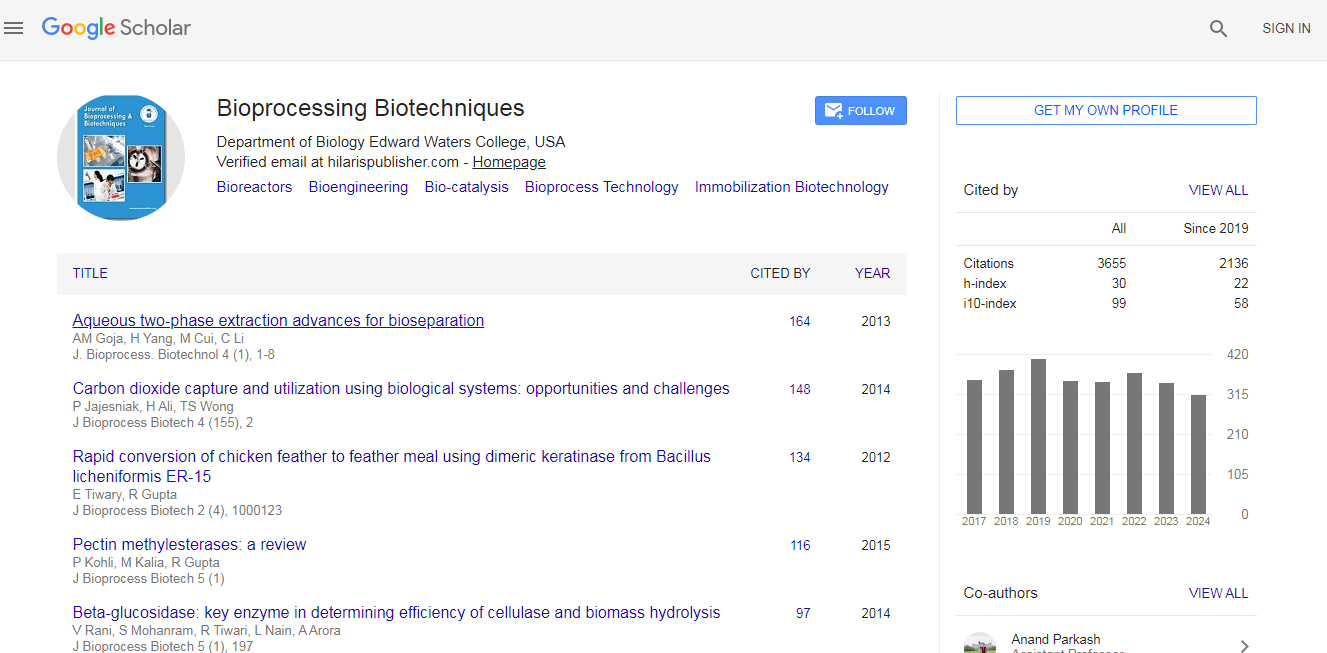
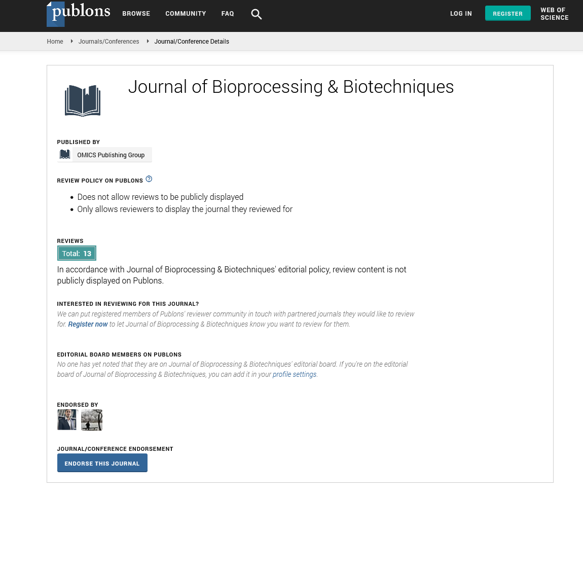
Citation: Mandalari, Giuseppina. “Commercial Food Microorganism Biofilm Communities.” J Bioprocess Biotech 12 (2022): 544.
Copyright: © 2022 Mandalari G. This is an open-access article distributed under the terms of the Creative Commons Attribution License, which permits unrestricted use, distribution, and reproduction in any medium, provided the original author and source are credited.
Biofilms, which exist as microorganisms on surfaces and can increase food cross contamination, can change the cleaning and disinfection dynamics in the food industry. Biofilm is an association of microorganisms that is irreversibly linked to a surface and is contained in an extracellular polymeric substance matrix, posing a significant challenge to the food industry. A strong disinfectant is required to eliminate bacterial attachments in order to prevent biofilm formation and eliminate them from the reversible attachment and irreversible stages, where attached microorganisms improve surface adhesion. This review paper addresses biofilm issues from all angles, including biofilmforming pathogens in the food industry, biofilm disinfectant resistance, and biofilm identification methods. Biofilms are thought to be responsible for food spoilage and outbreaks, as well as damage to food processing equipment [1].
Bacteria typically bind to surfaces and form spatially structured communities within a self-produced matrix made up of extracellular polymeric substances known as biofilms. Biofilms pose significant challenges for the food industry because they allow bacteria to bind to a variety of surfaces, including rubber, polypropylene, plastic, glass, stainless steel, and even food products, in a matter of minutes, with mature biofilms developing in a matter of days [2]. Since ancient times, this sessile life form has been regarded as an excellent survival technique for microorganisms, owing to the protective barrier generated and physiological changes caused by the biofilm matrix, while fighting against the adverse environmental circumstances commonly encountered by bacteria in man-made and natural settings, including food processing facilities.
Sensory tests that involve visually inspecting surfaces with good lighting, smelling unpleasant odours, and feeling encrusted or greasy surfaces are often performed as a process regulation to immediately overcome visible sanitation defects, whereas microbiological evaluations are frequently performed to ensure consistency with microbial standards and to improve sanitation procedures. It is well known that visual inspection does not correspond to bacterial counts. For all of the aforementioned reasons, the hygienic conditions of food-contact surfaces must be thoroughly examined. However, the lack of convergence among the various approaches used to detect and quantify biofilms makes it more difficult for the food industry to identify the most effective ones. The Hazard Analysis and Critical Control Points (HACCP) system, as well as good manufacturing practices, have been developed to address this issue [3].
The primary goals of this review were to identify the most significant biofilm examples in the food industry and to present methods for visualizing in situ biofilm production, avoiding this production, and removing biofilms. This study focuses on microbial biofilms in the food industry and provides an overview of their importance in cross-contamination when food comes into contact with surfaces. Although the goal of this work is not to go into detail in each discipline, specifically microbiology for biofilm isolation and identification.
Hydrophobicity, electrostatic charging, interface roughness, and topography of attachment surface affect biofilm formation and thus the overall hygiene status of the surface. Nonetheless, the precise outcome of some parameters varies greatly depending on the laboratory conditions. Some studies have found that rougher surfaces are more likely to support bacterial attachment, while others have found no link between roughness and bacterial attachment. Hydrophobic surfaces tend to attract more bacteria, but studies on the hydrophobicity effect show contradictory results, and other experiments show that hydrophilic surfaces allow for more bacterial adhesion than hydrophobic equivalents. The lack of clear results could be attributed to the various methods and bacterial strains used, as well as overall attachment being established for a variety of reasons [4].
Biofilm-forming species appear in factory environments in the food industry and can be pathogenic to humans due to the formation of biofilm structures. The food industry's processing environments, such as wood, glass, stainless steel, polyethylene, rubber, polypropylene, and so on, act as artificial substrates for these pathogens. When considering cleaning and disinfection processes, the characteristics of the bacterial growth form on food in a processing environment involve different behaviours. Controlling biofilm formations in the food industry can be difficult when determining the best strategy.
Biofilm bacteria have a distinct phenotype with a genotype in terms of gene transcription and growth rates under very specific conditions that differ from planktonic conditions. Biofilms can adhere to a wide range of surfaces with varying biotic and abiotic compositions, including human tissue and medical devices. Biofilms are a major threat once they form because they cause infectious diseases and economic loss. Several authors published additional research works on biofilm evolution and surface relations for marine microorganisms and seawater in the 1940s. Nonetheless, significant progress has been made since the introduction of the electron microscope, which enables high-resolution photomicroscopy at much higher magnifications than light microscopy.
Crystal violet staining measures the amount of dye incorporated into bacterial cell walls and is dependent on cell integrity but not viability. Other methods, such as ATP bioluminescence, report the cell's metabolic status, which drops to undetectable levels within minutes of cell death. Using both methods can provide additional information on the disinfectant-exposed cell. The findings suggest that, despite a significant drop in viable cell numbers in the biofilm following disinfectant treatment, a significant number of intact cells, or cellular debris, may still be capable of retaining the dye. This observation raises concerns about the dependability of crystal violet staining as a method of monitoring biofilm disinfection.
The use of fluorescent compounds to visualise a cell provides a wealth of information for analysing cell functions. Fluorescent compounds can be used to stain various activities and cell structures. Cell membranes, nucleotides, and proteins are the most common cell components. Depending on the molecule charge, hydrophobicity, or reactivity, the stain can enter cells. Small neutral and positively charged fluorescent compounds can thus normally reach mitochondria and dye them. Negatively charged molecules are unable to cross viable cell membranes. Because ester can pass through viable cell membranes and be hydrolyzed by cellular esterase’s into a negatively charged compound, it is an excellent functional group for staining viable cells [5].
The spatial resolution of an optical microscope is provided by this nondestructive analytical technique for fingerprint spectra. This novel technique allows for the quantitative, label-free, non-invasive, and rapid monitoring of biochemical changes in complex biofilm matrice. Raman spectra studies are distinguished by high specificity, revealing sharper, clearer bands than IR spectra, and a low water background. In contrast to IR microscopy, Raman spectroscopy can use visible light for excitation, allowing standard optics to be used.
None.
There are no conflicts of interest by author.
Google Scholar, Crossref, Indexed at
Google Scholar, Crossref, Indexed at
Google Scholar, Crossref, Indexed at
Google Scholar, Crossref, Indexed at
Journal of Bioprocessing & Biotechniques received 3351 citations as per Google Scholar report