Commentary - (2025) Volume 11, Issue 1
Received: 01-Feb-2025, Manuscript No. jotr-25-168443;
Editor assigned: 03-Feb-2025, Pre QC No. P-168443;
Reviewed: 15-Feb-2025, QC No. Q-168443;
Revised: 20-Feb-2025, Manuscript No. R-168443;
Published:
27-Feb-2025
, DOI: 10.37421/2476-2261.2025.11.295
Citation: Thomas, Vir liano. "Overexpression of MISP is found in Gastric Cancer and Intestinal Metaplasia." J Oncol Transl Res 11 (2025): 295.
Copyright: © 2025 Thomas V. This is an open-access article distributed under the terms of the Creative Commons Attribution License, which permits
unrestricted use, distribution, and reproduction in any medium, provided the original author and source are credited.
The global burden of cancer continues to rise, with over 19 million new cases and nearly 10 million cancer-related deaths reported in 2024 alone. This growing demand for effective oncology care has placed considerable pressure on healthcare systems, prompting the exploration of innovative technologies to enhance efficiency, accuracy, and personalization. Among these, Artificial Intelligence (AI) has emerged as a transformative force in clinical oncology. By leveraging machine learning algorithms, deep learning models, and big data analytics, AI has the potential to revolutionize every stage of cancer managementâ??from early detection and diagnosis to prognosis prediction, treatment planning, and response monitoring [1].
AI in oncology is not just about automation but about augmentationâ??enhancing the capabilities of clinicians through data-driven insights and predictive modeling. It enables the integration of vast, complex datasets including radiologic images, pathology slides, genomic data, and electronic health records (EHRs) to uncover patterns that may not be discernible through traditional approaches. As AI becomes more sophisticated and embedded in clinical workflows, it holds the promise of delivering more precise, timely, and personalized oncology care [2].
Medical imaging has long been a cornerstone of cancer detection and monitoring. AI-driven image analysis is now transforming radiology by enabling more accurate and efficient interpretation of imaging studies. AI-powered CAD systems assist radiologists in identifying malignant lesions in mammograms, chest CTs, and MRIs. Deep convolutional neural networks (CNNs) trained on thousands of annotated images can detect abnormalities with high sensitivity and specificity, often surpassing human readers in certain settings. Radiomics involves the extraction of quantitative features from imaging data, such as tumor shape, texture, and intensity. AI algorithms can analyze these features to distinguish between benign and malignant lesions, predict tumor aggressiveness, and guide biopsy decisions. For example, AI models using radiomic features have shown promise in differentiating indolent from aggressive prostate cancer on multiparametric MRI [3].
AI applications in screening programs, such as those for breast, lung, and colorectal cancer, can improve detection rates and reduce false positives. Google's AI model for mammography interpretation, for instance, demonstrated greater accuracy than radiologists in a landmark study. Histopathology remains the gold standard for cancer diagnosis. AI is revolutionizing pathology by enabling digital slide analysis, thereby enhancing diagnostic accuracy and reducing interobserver variability. AI models can evaluate WSI to identify cancer subtypes, grade tumors, and detect micrometastases in lymph nodes. AI tools like PathAI and Paige.AI use deep learning to classify tumors based on morphological features, improving diagnostic precision. AI can predict molecular alterations (e.g., EGFR mutations, HER2 status) directly from histology images, offering a non-invasive alternative to genetic testing. AI enhances the interpretation of liquid biopsiesâ??blood tests that detect circulating tumor DNA (ctDNA), exosomes, or microRNAsâ??by identifying subtle genomic patterns and distinguishing cancer-specific signals from background noise. This approach holds promise for early detection, minimal residual disease monitoring, and recurrence prediction [4].
Effective cancer management requires ongoing assessment of treatment response and early detection of recurrence. AI aids in these processes through. AI algorithms can quantitatively assess changes in tumor volume and characteristics on imaging, enabling earlier identification of non-responders. AI-powered platforms track symptoms, vital signs, and quality-of-life metrics using wearable sensors and mobile apps, providing clinicians with real-time data on treatment impact. Machine learning can anticipate treatment-related toxicities (e.g., neutropenia, cardiotoxicity) based on baseline characteristics and real-time monitoring data [5].
Google Scholar Cross Ref Indexed at
Google Scholar Cross Ref Indexed at
Google Scholar Cross Ref Indexed at
Google Scholar Cross Ref Indexed at
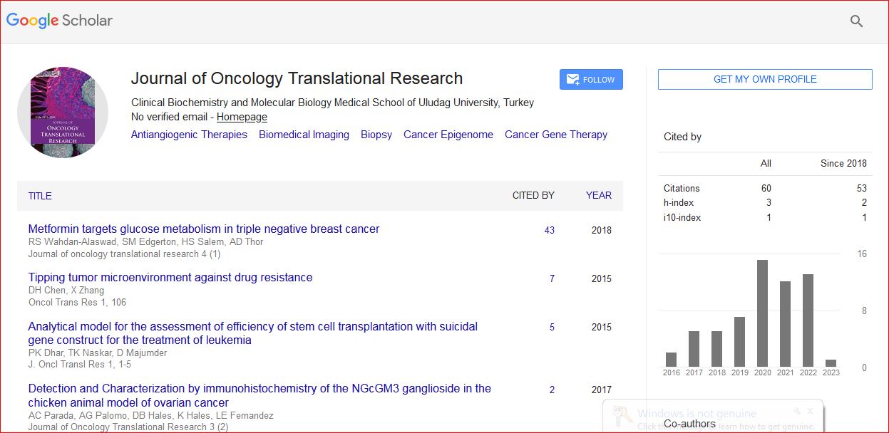
Journal of Oncology Translational Research received 93 citations as per Google Scholar report