Margaret Simonian, Marcus A Stoodley and Mark P Molloy
Scientific Tracks Abstracts: J Comput Sci Syst Biol
Identification of membrane proteins that are expressed on the arteriovenous malformation (AVM) endothelium post radiosurgery is of fundamental importance in developing a new treatment of brain AVMs. Arteriovenous malformations are tangle of abnormal arteries and veins referred to as nidus of AVM, linked by one or more fistula. They are the most common cause of haemorrhagic stroke in children and young adults. Radiosurgery is the treatment option recommended for lesions less than 3cm in diameter and located in eloquent area where surgery can cause neurological defect. However vascular occlusion after radiosurgery can take up to 3 years to complete, during that time patients remain at risk of haemorrhage. This study aims to identify unique protein targets in AVM endothelium that are different from normal vessels post radiosurgery. These proteins can then be used as targets for a ligand-directed vascular treatment to promote rapid thrombosis in AVM vessels. In vitro biotinylation and iTRAQ mass spectrometry was used to assess membrane protein changes in the murine endothelial cells (bEnd.3) post radiation. bEnd.3 cell cultures were irradiated with 25Gy and surface biotinylation was performed at 6h, 24h, 48h and 72 hours post radiation. Biotinylated proteins were captured on streptavidin resin, digested with trypsin, then labelled with iTRAQ reagent. Peptides were separated by SCX and analysed by nanoLC/ MS. iTRAQ-MS of biotinylated proteins in bEnd.3 cells detected on average 112 proteins from three independent biological replicates with 95% confidence. Eleven proteins were significantly differentially expressed in at least two out of three experiments. The most extensive changes were observed after 48h post radiation. Two known membrane proteins, CD109 and cadherin 5 were regulated after radiation. The other nine proteins are not commonly expected to be cell surface localised, but appeared following radiation. Immunocytochemistry of cell cultures was also conducted to confirm the localisation and expression of these molecules. Currently these proteins are being validated with MSE. Preliminary use of in vivo biotinylation in a rat model of AVM was also carried out and proteins identified using the method described above. The rat was perfused with NHS-LS-biotin, the fistula was harvested, membrane proteins were extracted and captured on streptavidin resin then digested overnight with trypsin and analysed by nano LC/MS. This preparation contained 135 proteins, 16 of them were known membrane proteins. Examples included various integrins, CD markers, adhesion proteins and ATPases. Further optimisation of the in vivo labelling protocol will be carried out to examine parameters such as perfusion time and biotin concentration. Samples will then be assessed by nanoLC/MS. In conclusion, Cell surface protein biotinylation and iTRAQ mass spectrometry of murine endothelial cells identified protein targets in response to irradiation.
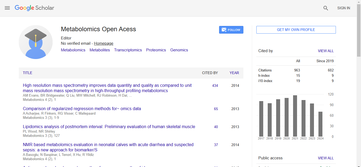
Metabolomics:Open Access received 895 citations as per Google Scholar report

 Spanish
  Chinese
  Russian
  German
  French
  Japanese
  Portuguese
  Hindi
Hindi 