Perspective - (2025) Volume 9, Issue 4
Received: 01-Jul-2025, Manuscript No. jigc-26-185922;
Editor assigned: 03-Jul-2025, Pre QC No. P-185922;
Reviewed: 17-Jul-2025, QC No. Q-185922;
Revised: 22-Jul-2025, Manuscript No. R-185922;
Published:
29-Jul-2025
, DOI: 10.37421/2684-4591.2025.9.331
Citation: Larsen, Henrik. ”AI Real-Time Plaque Elasticity for
Fluoroless Angioplasty.” J Interv Gen Cardiol 09 (2025):331.
Copyright: © 2025 Larsen H. This is an open-access article distributed under the terms of the Creative Commons Attribution License, which permits unrestricted use,
distribution and reproduction in any medium, provided the original author and source are credited.
The field of interventional cardiology is undergoing a transformative shift towards enhancing procedural guidance through advanced imaging and artificial intelligence (AI) [1].
The development of real-time AI-driven plaque elasticity indexing during fluoroless angioplasty represents a significant leap forward, aiming to provide immediate, quantitative feedback on atherosclerotic plaque properties without the use of ionizing radiation [1].
This novel approach seeks to improve lesion assessment and treatment selection, thereby making interventional procedures safer and more informative for both patients and clinicians [1].
Intravascular ultrasound (IVUS) and optical coherence tomography (OCT) are instrumental in characterizing atherosclerotic plaque, offering detailed anatomical and histological information that serves as the bedrock for developing quantitative elasticity measures [2].
The synergy between AI and these high-resolution imaging modalities is paramount for achieving real-time plaque assessment capabilities [2].
Across various medical disciplines, the application of artificial intelligence in medical imaging is experiencing rapid expansion, with deep learning techniques showing particular promise in cardiovascular imaging [3].
These AI applications span tasks such as lesion detection, segmentation, and risk stratification, highlighting the potential for automating complex analyses and delivering real-time insights that directly support methodologies like plaque elasticity indexing [3].
Concurrently, there is a notable trend towards fluoroless or reduced-fluoroscopy procedures in interventional cardiology, driven by concerns over radiation exposure [4].
This shift necessitates the development of sophisticated imaging and guidance systems to compensate for the absence of traditional fluoroscopic guidance, underscoring the relevance of techniques such as AI-based plaque elasticity indexing [4].
The mechanical properties of atherosclerotic plaques, particularly their elasticity, are critical determinants of plaque rupture risk and subsequent interventional outcomes [5].
Studies have demonstrated that stiffer plaques are often associated with higher rates of adverse clinical events, reinforcing the clinical utility of quantifying plaque elasticity to guide treatment decisions effectively [5].
Beyond pressure wire measurements, the assessment of hemodynamic significance is being advanced through techniques that integrate imaging data, such as computational fluid dynamics [6].
This exploration underscores a growing demand for multi-parametric assessments in interventional guidance, where plaque elasticity can contribute meaningfully to a comprehensive evaluation [6].
Machine learning models are increasingly being developed to predict cardiovascular events by leveraging imaging biomarkers, with deep learning approaches showing considerable success [7].
These models can extract intricate patterns from imaging data, such as IVUS, that correlate with clinical outcomes, a principle directly applicable to the development and refinement of plaque elasticity indexing methods [7].
For complex percutaneous coronary interventions (PCI), real-time guidance is crucial, and advanced imaging technologies are at the forefront of improving outcomes [8].
The integration of AI with these technologies further enhances their utility, emphasizing the need for quantitative, real-time metrics like plaque elasticity to guide device selection and deployment effectively [8].
The feasibility of deriving plaque stiffness from imaging data using non-invasive techniques is being explored, offering a promising pathway for characterization [9].
Research in this area supports the foundational principles required for the development of AI-driven elasticity estimation methods from readily available imaging modalities [9].
Finally, the successful integration of AI into real-time clinical decision support systems in the cardiac catheterization laboratory is a significant trend, requiring careful consideration of data integration, validation, and user interface design [10].
AI tools that deliver actionable information, such as plaque elasticity measurements, are vital for empowering interventional cardiologists to make informed decisions during procedures [10].
The core of this research lies in the proposal of real-time AI-driven plaque elasticity indexing during fluoroless angioplasty, a technique designed to significantly enhance procedural guidance [1].
By offering immediate, quantitative insights into the mechanical properties of atherosclerotic plaques, this approach aims to overcome the limitations of traditional methods and provide clinicians with a more comprehensive understanding of lesion characteristics without resorting to ionizing radiation [1].
The ultimate goal is to refine lesion assessment and optimize treatment selection, thereby improving the overall safety and efficacy of interventional cardiology procedures [1].
Central to the development of accurate and reliable plaque elasticity measures are advanced intravascular imaging modalities such as intravascular ultrasound (IVUS) and optical coherence tomography (OCT) [2].
These technologies furnish exceptionally detailed anatomical and histological information about atherosclerotic plaques, which is indispensable for establishing the quantitative elasticity metrics proposed by this work [2].
The seamless integration of artificial intelligence with these sophisticated imaging techniques is a critical enabler for real-time plaque assessment [2].
The pervasive adoption of artificial intelligence in medical imaging is a hallmark of modern healthcare, with deep learning algorithms at the forefront of innovation in cardiovascular imaging [3].
These AI applications are being leveraged for a multitude of tasks, including the precise detection and segmentation of lesions, as well as the stratification of cardiovascular risk [3].
The capacity of AI to automate complex analytical processes and deliver real-time, actionable insights directly aligns with and supports the development of AI-driven plaque elasticity indexing [3].
The ongoing transition towards fluoroless or significantly reduced-fluoroscopy interventional cardiology procedures is motivated by the imperative to minimize patient and operator radiation exposure [4].
This paradigm shift necessitates the invention and implementation of advanced imaging and guidance systems that can effectively substitute for conventional fluoroscopic visualization [4].
Consequently, the development of novel techniques, such as AI-based plaque elasticity indexing, becomes critically important to provide the necessary guidance in these radiation-sparing procedures [4].
The mechanical characteristics of atherosclerotic plaques, particularly their elasticity, are profoundly influential factors in determining the risk of plaque rupture and the subsequent outcomes of interventional procedures [5].
Empirical evidence consistently indicates that plaques exhibiting increased stiffness are associated with a higher incidence of adverse clinical events [5].
This correlation strongly supports the clinical value and necessity of accurately quantifying plaque elasticity as a means to inform and guide therapeutic interventions [5].
In the realm of interventional guidance, the assessment of hemodynamic significance is evolving beyond traditional methods like pressure wire measurements [6].
Emerging techniques, including the derivation of computational fluid dynamics from imaging data, are contributing to a more comprehensive understanding of coronary artery hemodynamics [6].
This progress highlights an increasing need for multi-parametric assessments, where the contribution of plaque elasticity to a holistic evaluation of lesion severity and risk is likely to be significant [6].
The predictive power of machine learning models for cardiovascular events, when trained on imaging biomarkers, is a rapidly advancing area of research [7].
Specifically, deep learning approaches have demonstrated remarkable success in utilizing data from modalities like IVUS to predict plaque progression and the risk of rupture [7].
The underlying principle of AI's ability to discern complex patterns in imaging data that correlate with clinical outcomes is directly applicable to the task of developing robust plaque elasticity indexing methods [7].
For intricate percutaneous coronary interventions (PCI), the availability of real-time guidance is paramount for achieving optimal procedural outcomes [8].
Current and emerging imaging technologies, especially when augmented by AI, are pivotal in this regard [8].
There is a clear and present need for quantitative, real-time metrics that can assist interventionalists in making critical decisions concerning device selection and optimal deployment strategies, a need that plaque elasticity indices are well-positioned to address [8].
The exploration of atherosclerotic plaque mechanical properties using non-invasive techniques, and their subsequent correlation with established imaging features, provides a strong foundation for this work [9].
This research has demonstrated that plaque stiffness can be effectively estimated from medical imaging data, paving the way for non-invasive characterization [9].
Such findings lend significant credibility to the feasibility and potential of AI-driven elasticity estimation from readily available imaging sources [9].
Finally, the effective integration of artificial intelligence into real-time clinical decision support systems within the cardiac catheterization laboratory is a critical ongoing development [10].
This integration presents both challenges and opportunities related to data management, validation protocols, and the design of intuitive user interfaces [10].
The development of AI tools that furnish interventionalists with directly actionable information, such as precise plaque elasticity measurements, is essential for enhancing clinical decision-making and improving patient care [10].
This research proposes a novel method for real-time AI-driven plaque elasticity indexing during fluoroless angioplasty to improve interventional cardiology guidance. The technique aims to provide immediate quantitative feedback on atherosclerotic plaque properties without ionizing radiation, enhancing lesion assessment and treatment selection. It leverages advanced intravascular imaging modalities like IVUS and OCT, integrating them with AI and deep learning to extract detailed plaque characteristics. This approach addresses the growing need for radiation-sparing procedures and provides a crucial metric for assessing plaque vulnerability and rupture risk. The development is supported by research showing the correlation between plaque stiffness and adverse events, and the feasibility of estimating mechanical properties from imaging data. Ultimately, this AI-driven tool seeks to offer actionable insights for interventionalists in real-time decision support systems within the cardiac catheterization laboratory.
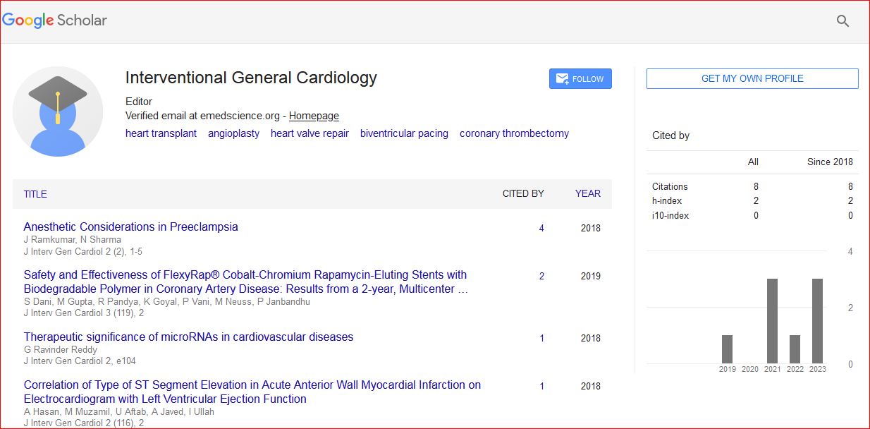
Journal of Interventional and General Cardiology received 11 citations as per Google Scholar report