Opinion - (2023) Volume 14, Issue 1
Received: 30-Jan-2023, Manuscript No. hgec-23-101983;
Editor assigned: 01-Feb-2023, Pre QC No. P-101983;
Reviewed: 15-Feb-2023, QC No. Q-101983;
Revised: 20-Feb-2023, Manuscript No. R-101983;
Published:
27-Feb-2023
, DOI: 10.37421/2161-0436.2023.14.197
Citation: Dcudo, Flaria. “The Embryo: Unveiling the Wonders of
Early Life Development.” Human Genet Embryol 14 (2023): 197.
Copyright: © 2023 Dcudo F. This is an open-access article distributed under the terms of the Creative Commons Attribution License, which permits unrestricted use, distribution, and reproduction in any medium, provided the original author and source are credited.
The embryo is a phase in the early development of multicellular organisms, representing a crucial stage in the journey from conception to the formation of a fully developed organism. During this period, the embryo undergoes remarkable transformations, laying the foundation for the complex structures and systems that will define its future existence. In this essay, we will explore the wonders of the embryo, delving into its formation, growth, and the intricate processes that shape its development. The journey of the embryo begins with the miraculous event of fertilization. Fertilization occurs when a sperm cell successfully penetrates and fuses with an egg, resulting in the formation of a zygote—the first cell of the new organism. The zygote contains the combined genetic material from both parents, providing the blueprint for the development of the entire organism. As the number of cells increases, the embryo transforms into a structure known as a blastocyst. The blastocyst is a hollow sphere composed of two distinct cell populations: the outer layer called the trophoblast and the inner cell mass. The trophoblast plays a crucial role in implantation the process by which the embryo attaches to and embeds itself into the uterine wall. Meanwhile, the inner cell mass gives rise to the embryo proper and is responsible for the development of all major body structures [1].
Endometrial As implantation occurs, the embryo continues its remarkable development. The inner cell mass differentiates into three primary germ layers: the ectoderm, mesoderm, and endoderm. These germ layers serve as the foundation for the development of all organs, tissues, and systems in the body. The ectoderm, the outermost germ layer, gives rise to the nervous system, including the brain and spinal cord, as well as the skin and its appendages. The mesoderm, situated between the ectoderm and endoderm, develops into various structures such as muscles, bones, blood vessels, connective tissues, and the urogenital system. Finally, the endoderm, the innermost layer, forms the lining of the digestive and respiratory tracts, as well as certain glands. The process of differentiation and specialization within the germ layers is guided by an intricate interplay of genetic and environmental factors.
As the embryo progresses in development, it undergoes a process called morphogenesis— the shaping and organization of tissues and structures to create the three-dimensional architecture of the body. Morphogenesis involves a variety of cellular processes, including cell migration, proliferation, differentiation, and changes in cell shape. These processes work in harmony to sculpt and position organs, form complex structures, and establish connections between cells. During morphogenesis, the embryo goes through a series of critical events. Neurulation involves the bending and fusion of specialized cells along the dorsal midline of the embryo, resulting in the formation of the brain and spinal cord. In addition to morphogenesis, another essential process in embryo development is organogenesis—the formation of organs. Organogenesis involves the precise coordination of cellular interactions, tissue growth, and differentiation to create the various organs and structures that will function collectively in the mature organism [2,3].
The embryo is exquisitely sensitive to external influences during its developmental journey. Exposure to certain substances, such as drugs, alcohol, or toxins, can have detrimental effects on the developing embryo and may lead to congenital abnormalities or developmental disorders. Therefore, ensuring a healthy and supportive environment for the embryo is of utmost importance for its proper growth and development. As the embryo progresses through its developmental stages, it eventually transitions into the next phase known as the fetus. The transition from embryo to fetus is marked by the formation of the major organ systems, increased differentiation of tissues, and the development of distinct human characteristics. During the fetal stage, growth and refinement of structures and systems continue, preparing the organism for the eventual transition to life outside the womb [4,5].
In conclusion, the embryo represents an awe-inspiring stage in the early development of multicellular organisms. From the moment of fertilization, the embryo undergoes a remarkable series of transformations, culminating in the formation of complex organs and structures. Through the processes of cleavage, blastocyst formation, germ layer differentiation, morphogenesis, and organogenesis, the embryo establishes the foundation for the development of a fully functional organism. The intricate interplay between genetic and environmental factors guides the embryo's growth and ensures the formation of the diverse tissues and organs that make up the human body. Understanding the wonders of embryo development not only deepens our appreciation for the complexity of life but also provides valuable insights into the prevention and treatment of developmental disorders and birth defects.
None.
None.
Google Scholar, Crossref, Indexed at
Google Scholar, Crossref, Indexed at
Google Scholar, Crossref, Indexed at
Google Scholar, Crossref, Indexed at
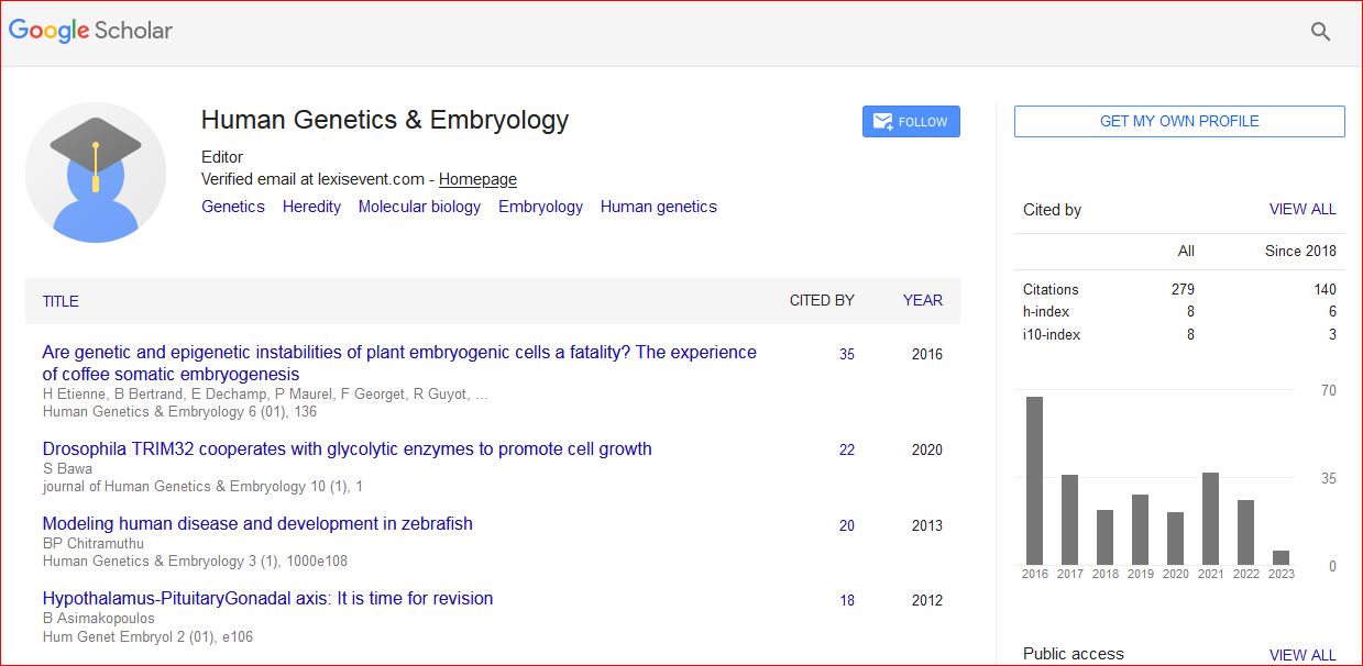
Human Genetics & Embryology received 309 citations as per Google Scholar report