Case Report - (2026) Volume 16, Issue 1
Received: 01-Dec-2025, Manuscript No. jccr-25-175710;
Editor assigned: 02-Feb-2026, Pre QC No. P-175710;
Reviewed: 04-Feb-2026, QC No. Q-175710;
Revised: 12-Feb-2026, Manuscript No. R-175710;
Published:
19-Feb-2026
, DOI: 10.37421/2165-7920.2026.16.1699
Citation: Shirvani, Masoud, Mohammadali Arami, Behrang Kazeminezhad and Zeynab. Kishani Farahani. “Rapidly Progressive Paraplegia in Intramedullary Thoracic Ependymoma: Evidence for Prompt Surgical Intervention (Case Report).” J Clin Case Rep 16 (2026): 1699.
Copyright: © 2026 Shirvani M, et al. This is an open-access article distributed under the terms of the Creative Commons Attribution License, which permits unrestricted use, distribution, and reproduction in any medium, provided the original author and source are credited.
Intramedullary spinal ependymomas often grow slowly and are frequently referred for surgery on a non-urgent, routine basis. Patients may also lose time consulting with surgeons and making decisions and in the meantime may develop irreversible disabilities.
In this report, we present a patient who developed rapid paraplegia, highlighting the need for early surgery for intramedullary ependymomas.
The most common intramedullary tumors of the spinal cord are astrocytoma and ependymoma, with ependymoma being the most frequent intramedullary tumor in adults [1]. Clinical signs of intramedullary spinal cord tumors, including ependymoma, typically include paresthesia in the limbs, gradually progressive weakness, pain in the corresponding spinal segments and sphincter dysfunction. Symptoms usually begin mildly and worsen over months [2-4]. Also, thoracic spinal cord ependymomas usually appear to be more aggressive [5].
When symptoms begin acutely and progress to complete paralysis in a fulminant manner, diagnoses such as transverse myelitis, Guillain–Barré syndrome, hematomyelia, extramedullary compressive lesions and infections such as epidural abscess are often considered and the necessary investigations and treatments are undertaken [6]. Delay in diagnosis and treatment in these cases can lead to severe and permanent disability.
In contrast, such a history and course are unusual and rare for ependymomas. There are only a few reports in the literature in which acute paraplegia has been attributed to intramedullary hemorrhage [7, 8]. Here we present a case that presents with rapidly progressive paraplegia without intralesional hemorrhage, highlighting important lessons for surgeons in the management of these patients.
A 57-year-old woman presented to her family physician with upper back pain. On neurological examination performed by the family physician, the strength of all four limbs was normal. She did not complain of numbness or tingling in the limbs. Balance and gait tests were normal. She had no local tenderness. There was no history of trauma. She did not complain of sphincter dysfunction.
Based on the examination and history, myofascial pain was diagnosed and the patient was prescribed physiotherapy and local analgesic treatments.
The patient's pain did not improve and the patient returned with complaints of numbness in the limbs and slight imbalance. The patient was referred to a neurosurgeon for further evaluation. On examination, there was increased tendon reflexes and weakness in the lower limbs of 4.5 was discovered. An emergency MRI of the cervical and thoracic spine was requested.
An intramedullary lesion with spinal cord swelling was observed on MRI, which was enhanced by gadolinium injection (Figure 1). On physical examination, which was performed approximately 24 hours after the first neurosurgeon's examination, the weakness of the limbs had worsened and the patient was able to walk only with assistance. The patient was referred to our specialized neurosurgical center for surgery and unfortunately, within 24 hours, the patient's weakness had decreased to 1.5 and urine and fecal incontinence. The patient underwent surgery. After laminectomy and myelotomy, the tumor was completely removed. Contrary to our initial suspicion, no signs of any hemorrhage were seen. The patient was discharged for rehabilitation measures and followed up.
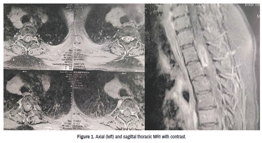
Figure 1. Axial (left) and sagittal thoracic MRI with contrast.
Pathological examination
Microscopic evaluation of the cellular tumor reveals infiltrative mass with monomorphic round to oval cells with speckled chromatin arranged in perivascular pseudorosettes and true ependymal rosettes, lumina and fibrillar areas.
In IHC study, the tumor was positive for S100, GFAP, vimentin, perinuclear dot-like pattern of EMA staining. CD56 staining in lumina and tumor cells was also seen. Unexpectedly tumor had high mitotic activity. As for molecular study N_Myc amplification returned negative.
Spinal cord ependymoma is a relatively common intramedullary tumor across a broad age range. The clinical significance of this tumor is well established, making timely diagnosis and treatment particularly important, as progressive disease can lead to permanent symptoms and substantial disability.
Advances in imaging technologies have improved early diagnosis, often before significant disability has occurred. The indolent growth of many spinal ependymomas typically permits surgical intervention at an appropriate time, allowing patients to resume life with minimal or no disability.
However, the case presented here demonstrates that this course is not universal. Tumor growth and symptom progression are not invariably slow or gradual; they can evolve over months to years and may, in some instances, present acutely. Consequently, prompt surgical intervention is essential. Patients should not be placed on prolonged waiting lists for surgery but should be promptly referred to specialized centers with sufficient experience and resources. This experience informs the recommendation to our colleagues.
Although prior reports in the literature have suggested that hemorrhage may underlie rapid clinical deterioration [4], our case illustrates that ependymomas can present with fulminant symptoms even in the absence of bleeding. As our pathology colleagues showed, this tumor exhibited higher mitotic activity. Therefore, this feature could also explain the rapid progression of symptoms and signs.
Once a spinal intramedullary ependymoma is diagnosed, complete surgical resection should be considered at the earliest opportunity, because irreversible neurological deficits may occur rapidly.
None.
None.
Google Scholar, Cross Ref, Indexed at
Google Scholar, Cross Ref, Indexed at
Google Scholar, Cross Ref, Indexed at
Google Scholar, Cross Ref, Indexed at
Google Scholar, Cross Ref, Indexed at
Google Scholar, Cross Ref, Indexed at
Journal of Clinical Case Reports received 1345 citations as per Google Scholar report