Opinion - (2025) Volume 9, Issue 5
Received: 01-Sep-2025, Manuscript No. hps-26-184484;
Editor assigned: 03-Sep-2025, Pre QC No. P-184484;
Reviewed: 17-Sep-2025, QC No. Q-184484;
Revised: 22-Sep-2025, Manuscript No. R-184484;
Published:
29-Sep-2025
, DOI: 10.37421/2573-4563.2025.9.366
Citation: Popescu, Daniela S.. ”Non-invasive Liver Fibrosis: Biomarkers, Imaging, and Future Directions.” J Hepatol Pancreat Sci 09 (2025):366.
Copyright: © 2025 Popescu S. Daniela This is an open-access article distributed under the terms of the Creative Commons Attribution License, which permits unrestricted use,distribution and reproduction in any medium, provided the original author and source are credited.
The imperative to discover non-invasive biomarkers for the early detection of liver fibrosis is paramount for enhancing patient outcomes and improving the management of liver diseases. Traditional diagnostic methods, such as liver biopsy, are inherently invasive, carrying risks and limitations that hinder frequent monitoring and widespread application. Consequently, a significant paradigm shift is underway, focusing on the identification and validation of circulating molecules, advanced imaging techniques, and even genetic signatures that can reliably indicate the presence and severity of liver fibrosis without the need for tissue sampling. This evolution promises more accessible and frequent monitoring strategies, thereby enabling timely therapeutic interventions and potentially preventing the progression to more severe conditions like cirrhosis and hepatocellular carcinoma [1].
Within this evolving landscape, circulating microRNAs (miRNAs) have emerged as promising early indicators of liver fibrosis. These small, non-coding RNA molecules play fundamental roles in regulating gene expression, and their altered profiles in the context of liver disease can be detected in peripheral blood. Research efforts have successfully identified specific miRNA signatures associated with various stages of fibrosis, suggesting their significant utility as non-invasive diagnostic tools. However, a key challenge remains in validating these findings across diverse patient populations and in standardizing the detection methodologies to ensure reproducibility and clinical applicability [2].
Elastography, encompassing techniques such as transient elastography (TE) and magnetic resonance elastography (MRE), has established itself as a significant non-invasive modality for assessing liver stiffness, which serves as a direct surrogate marker for the degree of fibrosis. These imaging-based techniques provide quantitative measurements that have demonstrated strong correlations with histological staging of fibrosis. Current research is actively focused on refining the accuracy of elastography, particularly in distinguishing advanced fibrosis from cirrhosis, and on integrating these powerful tools more effectively into routine clinical practice for the comprehensive management of chronic liver diseases [3].
Extracellular matrix (ECM) components, including crucial proteins like collagen, laminin, and hyaluronic acid, are intrinsically involved in the complex processes of liver fibrosis. The development of novel biomarkers is actively pursuing the detection of elevated levels or specific fragments of these ECM proteins within the circulation. Assays designed to measure specific epitopes or degradation products of collagen, for example, show considerable promise in reflecting ongoing fibrogenesis, thereby offering a direct, non-invasive window into the dynamic process of scar tissue formation within the liver [4].
A particularly promising strategy for enhancing the diagnostic accuracy of non-invasive liver fibrosis assessment involves the integration of multiple biomarkers into composite scores or algorithms. By synergistically combining data derived from blood tests, advanced imaging modalities, and relevant clinical parameters, these multi-analyte approaches can construct a more comprehensive and nuanced picture of liver health. Such carefully curated panels aim to overcome the inherent limitations of individual markers and provide more precise staging of fibrosis, ultimately guiding more effective and personalized treatment decisions [5].
Genetic and epigenetic factors play a notable role in explaining the inter-individual variability observed in the progression of liver fibrosis. Investigating specific genetic polymorphisms within genes known to be involved in fibrogenesis, inflammation, and resolution pathways can effectively identify individuals who are at a higher risk of developing severe liver fibrosis. Concurrently, epigenetic modifications, such as alterations in DNA methylation patterns, may also serve as novel biomarkers that reflect the dynamic changes occurring in the cellular environment of fibrotic livers, opening avenues for personalized risk stratification [6].
The intricate relationship between the gut microbiome and the pathogenesis and progression of liver fibrosis is gaining increasing recognition within the scientific community. Dysbiosis, characterized by an imbalance in the composition and function of gut microbial communities, can lead to compromised intestinal barrier integrity and increased translocation of microbial products, thereby fueling liver inflammation and fibrogenesis. Furthermore, metabolites produced by gut bacteria, including short-chain fatty acids and modified bile acids, are also implicated in these processes. Profiling the microbiome and its metabolic output presents an exciting opportunity for the development of novel non-invasive biomarkers [7].
Proteomic approaches represent a powerful and versatile methodology for the identification of novel protein biomarkers intimately associated with the development and progression of liver fibrosis. By comprehensively analyzing the complete set of proteins expressed by cells or present in biological fluids, researchers are uncovering subtle yet significant changes that are indicative of fibrotic processes. The application of advanced mass spectrometry techniques is increasingly enabling the identification of well-defined panels of proteins capable of differentiating fibrotic stages with remarkable accuracy, moving beyond the limitations of single-target analysis [8].
Beyond conventional ultrasound and computed tomography (CT) scans, advanced imaging techniques are being rigorously explored for their capacity to detect and quantify liver fibrosis non-invasively. Modalities such as diffusion-weighted imaging (DWI) and contrast-enhanced ultrasound (CEUS) are capable of providing valuable information regarding tissue microstructure and vascularity, both of which are significantly altered during the fibrotic process. The development of quantitative imaging biomarkers holds substantial promise for enabling real-time assessment and precise monitoring of disease progression in a non-invasive manner [9].
Ultimately, the identification of novel non-invasive biomarkers for the early detection of liver fibrosis is a critical endeavor that facilitates early therapeutic intervention and significantly improves patient management strategies. While existing markers demonstrate considerable promise, ongoing research into circulating nucleic acids (including DNA and RNA), extracellular vesicles, and sophisticated advanced imaging technologies continues to expand the diagnostic armamentarium. The overarching goal remains the development of cost-effective, highly accurate, and readily accessible tools that can either replace or effectively supplement the role of liver biopsy in the routine assessment of liver fibrosis [10].
The pursuit of non-invasive biomarkers for early liver fibrosis detection is fundamentally driven by the need to improve patient outcomes. Traditional diagnostic methods, such as liver biopsy, are invasive and present inherent limitations, making them unsuitable for routine or frequent monitoring. This has spurred a significant research focus on alternative approaches, including the analysis of circulating molecules, the application of advanced imaging techniques, and the exploration of genetic signatures that can reliably indicate the presence and severity of fibrosis without requiring tissue sampling. This paradigm shift aims to facilitate more accessible and frequent monitoring, enabling timely interventions and potentially preventing the progression of liver disease to cirrhosis and hepatocellular carcinoma [1].
Among the promising circulating biomarkers, microRNAs (miRNAs) are gaining attention as early indicators of liver fibrosis. These small RNA molecules are involved in regulating gene expression, and their altered levels in the blood can reflect the state of liver disease. Studies have identified specific miRNA profiles associated with different fibrosis stages, suggesting their potential as non-invasive diagnostic tools. Key challenges in this field include the need for validation in diverse patient populations and the standardization of detection methods to ensure reliable and reproducible results [2].
Elastography techniques, particularly transient elastography (TE) and magnetic resonance elastography (MRE), represent significant advancements in the non-invasive assessment of liver stiffness, a direct indicator of fibrosis. These methods offer quantitative measurements that correlate well with histological fibrosis staging. Current research efforts are concentrated on enhancing the accuracy of elastography, especially in differentiating advanced fibrosis from cirrhosis, and on integrating these techniques seamlessly into the routine clinical management of chronic liver diseases [3].
Novel biomarkers are being developed to detect elevated levels or specific fragments of extracellular matrix (ECM) components, such as collagen, laminin, and hyaluronic acid, which are integral to liver fibrosis. Assays that measure specific epitopes or degradation products of collagen, for instance, show promise in reflecting active fibrogenesis. This approach provides a direct, non-invasive insight into the dynamic process of scar tissue formation in the liver [4].
To improve the accuracy of non-invasive liver fibrosis assessment, a strategy involving the integration of multiple biomarkers into composite scores or algorithms is being pursued. By combining data from blood tests, imaging, and clinical parameters, these multi-analyte approaches provide a more comprehensive evaluation of liver health. Such panels are designed to overcome the limitations of individual markers and offer more precise staging of fibrosis, thereby improving the guidance of treatment decisions [5].
Genetic and epigenetic factors contribute to the varying rates of liver fibrosis progression among individuals. Research into genetic polymorphisms in genes related to fibrogenesis, inflammation, and resolution pathways can help identify individuals at higher risk for severe fibrosis. Furthermore, epigenetic modifications, such as DNA methylation, may serve as novel biomarkers reflecting the altered cellular environment in fibrotic livers, paving the way for personalized risk stratification [6].
The influence of the gut microbiome on the pathogenesis and progression of liver fibrosis is increasingly recognized. Gut dysbiosis, an imbalance in microbial communities, can lead to increased intestinal permeability and the translocation of microbial products, promoting liver inflammation and fibrogenesis. Metabolites produced by gut bacteria, such as short-chain fatty acids and bile acids, are also implicated. Profiling the microbiome and its metabolic output holds potential for identifying novel non-invasive biomarkers [7].
Proteomic analysis offers a powerful method for identifying novel protein biomarkers associated with liver fibrosis. By examining the complete set of proteins in biological fluids or cells, researchers can detect subtle changes indicative of fibrotic processes. Advanced mass spectrometry techniques are facilitating the identification of protein panels that can accurately differentiate fibrotic stages, moving beyond single-analyte approaches [8].
Advanced imaging techniques, extending beyond standard ultrasound and CT, are being investigated for their ability to non-invasively assess and quantify liver fibrosis. Techniques like diffusion-weighted imaging (DWI) and contrast-enhanced ultrasound (CEUS) provide insights into tissue microstructure and vascularity, which are altered in fibrosis. The development of quantitative imaging biomarkers is crucial for real-time assessment and monitoring of disease progression [9].
The development of novel non-invasive biomarkers for early liver fibrosis detection is critical for timely intervention and improved patient management. While current markers show promise, ongoing research into circulating nucleic acids, extracellular vesicles, and advanced imaging continues to expand the diagnostic toolkit. The ultimate objective is to create cost-effective, accurate, and accessible tools that can replace or supplement liver biopsy for routine fibrosis assessment [10].
The field of liver fibrosis detection is shifting towards non-invasive methods due to the limitations of liver biopsy. Current research explores circulating molecules like microRNAs and extracellular matrix components, advanced imaging techniques such as elastography and DWI/CEUS, and proteomic profiling to identify novel biomarkers. Genetic and epigenetic factors are also being investigated for their role in fibrosis progression and risk stratification. The gut microbiome's influence on liver fibrosis is another area of active study. Integrating multiple biomarkers into composite scores shows promise for improved diagnostic accuracy. The ultimate goal is to develop accessible and accurate tools for early detection and management of liver fibrosis.
None
None
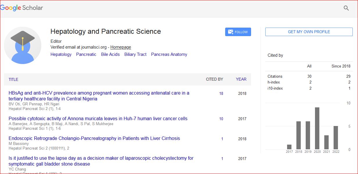
Hepatology and Pancreatic Science received 34 citations as per Google Scholar report