Commentary - (2025) Volume 10, Issue 3
Received: 01-May-2025, Manuscript No. JPNM-26-185775;
Editor assigned: 05-May-2025, Pre QC No. P-185775;
Reviewed: 19-May-2025, QC No. Q-185775;
Revised: 22-May-2025, Manuscript No. R-185775;
Published:
29-May-2025
, DOI: 10.37421/2472-100X.2025.10.347
Citation: Carter, Emily. ”Neuroimaging: Diagnosing Pediatric Brain Disorders.” J Pediatr Neurol Med 10 (2025):348.
Copyright: © 2025 Carter E. This is an open-access article distributed under the terms of the Creative Commons Attribution License, which permits unrestricted use, distribution and reproduction in any medium, provided the original author and source are credited.
Neuroimaging stands as a cornerstone in the diagnosis, characterization, and ongoing management of a wide spectrum of pediatric brain disorders. The ability of techniques such as Magnetic Resonance Imaging (MRI) and Computed Tomography (CT) to provide highly detailed anatomical information is absolutely essential for the accurate identification of structural abnormalities that may underlie various neurological conditions in children. These imaging modalities offer a non-invasive window into the developing brain, enabling clinicians to pinpoint deviations from typical development or identify pathological processes that might otherwise remain undetected. Beyond fundamental anatomical depiction, advanced MRI sequences have significantly expanded the diagnostic and research capabilities in pediatric neurology. Techniques like Diffusion Tensor Imaging (DTI) are invaluable for assessing the integrity of white matter tracts, which are crucial for inter-brain region communication and overall cognitive function. Similarly, functional MRI (fMRI) allows for the visualization of brain activity and connectivity patterns during cognitive tasks or rest states, offering profound insights into the dynamic functioning of the pediatric brain. These sophisticated neuroimaging tools are vital for understanding complex conditions such as autism spectrum disorder (ASD), where altered patterns of brain connectivity are increasingly recognized. They are also critical in the evaluation of epilepsy, developmental delays, and other neurodevelopmental disorders, providing objective data to support clinical assessments and guide therapeutic strategies. The early and accurate diagnosis facilitated by these technologies is a critical first step in improving long-term outcomes for affected children. The application of neuroimaging extends to the precise planning of interventions and the evaluation of their effectiveness over time. By providing objective measures of disease status and treatment response, these techniques allow for personalized treatment plans tailored to the individual child's needs. This individualized approach is particularly important in pediatric care, where developmental trajectories can vary significantly. Functional neuroimaging, with fMRI at the forefront, plays a particularly instrumental role in unraveling the complexities of aberrant brain activity observed in pediatric neuropsychiatric disorders. The insights gained from these studies are contributing to a more nuanced understanding of conditions such as Attention-Deficit/Hyperactivity Disorder (ADHD) and various anxiety disorders, paving the way for more targeted and effective interventions. Studies utilizing fMRI are actively revealing distinct patterns of functional connectivity within the brains of children diagnosed with these conditions. These patterns can differ significantly from those observed in typically developing children, offering potential biomarkers for diagnosis and prognostication. Such findings are crucial for differentiating between conditions that may present with overlapping symptoms. The ability to visualize how different brain regions communicate and interact in real-time provides a dynamic perspective that complements traditional structural assessments. This functional information is essential for understanding the underlying neurobiological mechanisms driving these complex disorders, highlighting the dynamic nature of brain function in the pediatric population. Diffusion tensor imaging (DTI) has indeed revolutionized the assessment of white matter development and integrity in pediatric populations. Its capacity to quantify the directionality and magnitude of water diffusion along axonal pathways allows for detailed evaluation of white matter microstructure. This is paramount for understanding the impact of various insults on the developing brain. DTI is particularly crucial for evaluating the effects of genetic disorders, perinatal injuries, and various neurodevelopmental conditions on the intricate network of white matter tracts. These injuries can disrupt axonal pathways, affecting the efficient transmission of neural signals and leading to a range of functional deficits. The objective measures provided by DTI aid in characterizing the extent of this damage. Furthermore, DTI enables the quantification of microstructural properties, such as fractional anisotropy and mean diffusivity, which serve as objective markers for tracking disease progression and assessing the efficacy of therapeutic interventions aimed at improving white matter health. This quantitative approach is essential for evidence-based clinical practice and the development of novel regenerative strategies.
Neuroimaging techniques are paramount in the diagnosis, characterization, and ongoing management of a diverse array of pediatric brain disorders. Modalities like MRI and CT offer unparalleled detailed anatomical information, which is fundamental for the identification of structural abnormalities that may contribute to various neurological conditions in children. These imaging methods provide a non-invasive view of the developing brain, assisting clinicians in pinpointing developmental deviations or pathological processes that might otherwise go unnoticed. Beyond basic anatomical visualization, sophisticated MRI sequences have substantially broadened the diagnostic and research capabilities within pediatric neurology. Techniques such as Diffusion Tensor Imaging (DTI) are indispensable for evaluating the integrity of white matter tracts, which are vital for inter-brain region communication and overall cognitive functioning. Likewise, functional MRI (fMRI) allows for the visualization of brain activity and connectivity patterns during cognitive tasks or periods of rest, yielding profound insights into the dynamic functioning of the pediatric brain. These advanced neuroimaging tools are critical for comprehending complex conditions like autism spectrum disorder (ASD), where altered patterns of brain connectivity are increasingly being recognized. They are also crucial in the assessment of epilepsy, developmental delays, and a host of other neurodevelopmental disorders, providing objective data to bolster clinical evaluations and guide therapeutic strategies. The early and accurate diagnosis facilitated by these technologies represents a critical initial step toward improving long-term outcomes for affected children. The application of neuroimaging extends significantly into the realm of precise intervention planning and the subsequent evaluation of treatment efficacy over time. By furnishing objective metrics of disease status and response to therapy, these techniques empower the creation of personalized treatment plans meticulously tailored to the unique needs of each child. This individualized approach is particularly salient in pediatric care, where developmental trajectories can exhibit considerable variability. Functional neuroimaging, with fMRI at its vanguard, plays an exceptionally instrumental role in elucidating the intricate mechanisms of aberrant brain activity observed in pediatric neuropsychiatric disorders. The insights gleaned from these investigations are contributing to a more sophisticated understanding of conditions such as Attention-Deficit/Hyperactivity Disorder (ADHD) and various anxiety disorders, thereby paving the way for the development of more targeted and effective interventions. Studies employing fMRI are actively uncovering distinct patterns of functional connectivity within the brains of children diagnosed with these conditions. These characteristic patterns can diverge significantly from those observed in typically developing children, potentially serving as biomarkers for diagnosis and prognosis. Such findings are instrumental in differentiating between disorders that may exhibit overlapping symptomatic presentations. The capability to visualize the real-time communication and interaction between different brain regions offers a dynamic perspective that complements traditional structural assessments. This functional information is indispensable for comprehending the underlying neurobiological mechanisms driving these complex disorders, underscoring the inherently dynamic nature of brain function in the pediatric population. Diffusion tensor imaging (DTI) has indeed ushered in a revolutionary era for the assessment of white matter development and integrity within pediatric cohorts. Its proficiency in quantifying the directionality and magnitude of water diffusion along axonal pathways facilitates a detailed evaluation of white matter microstructure, which is of paramount importance for understanding the impact of diverse insults on the developing brain. DTI holds particular significance in evaluating the repercussions of genetic disorders, perinatal injuries, and various neurodevelopmental conditions on the intricate architecture of white matter tracts. Such injuries can disrupt axonal pathways, thereby impeding the efficient transmission of neural signals and consequently leading to a spectrum of functional deficits. The objective data generated by DTI are crucial for precisely characterizing the extent of this damage. Moreover, DTI permits the quantitative assessment of microstructural properties, including fractional anisotropy and mean diffusivity, which serve as objective benchmarks for monitoring disease progression and gauging the effectiveness of therapeutic interventions designed to enhance white matter health. This quantitative methodology is indispensable for evidence-based clinical practice and the advancement of novel regenerative strategies.
Neuroimaging, encompassing techniques like MRI, CT, DTI, and fMRI, is critical for diagnosing, characterizing, and managing pediatric brain disorders. These methods provide detailed anatomical and functional insights into the developing brain, aiding in the identification of structural abnormalities and aberrant brain activity in conditions such as autism spectrum disorder, epilepsy, ADHD, and developmental delays. Advanced techniques like DTI assess white matter integrity, while fMRI reveals functional connectivity patterns. Neuroimaging is crucial for early detection, personalized treatment planning, and evaluating treatment efficacy, ultimately improving outcomes for children with neurological conditions. Its applications also extend to pediatric brain tumors, traumatic brain injuries, and genetic disorders, offering metabolic information through MRS and quantitative metrics for objective assessment.
None
None
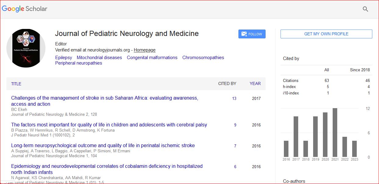
Journal of Pediatric Neurology and Medicine received 68 citations as per Google Scholar report