Brief Report - (2025) Volume 11, Issue 2
Received: 01-Mar-2025, Manuscript No. ldt-25-178409;
Editor assigned: 03-Mar-2025, Pre QC No. P-178409;
Reviewed: 17-Mar-2025, QC No. Q-178409;
Revised: 24-Mar-2025, Manuscript No. R-178409;
Published:
31-Mar-2025
, DOI: 10.37421/2472-1018.2025.11.295
Citation: Boateng, Samuel K.. ”Lung Inflammation: Drivers Of Acute and Chronic Injury.” J Lung Dis Treat 11 (2025):295.
Copyright: © 2025 Boateng K. Samuel This is an open-access article distributed under the terms of the Creative Commons Attribution License, which permits unrestricted use, distribution and reproduction in any medium, provided the original author and source are credited.
Inflammation is a fundamental process that plays a critical role in the development and progression of both acute and chronic lung injuries. In the context of acute lung injury, such as Acute Respiratory Distress Syndrome (ARDS), a rapid and severe inflammatory cascade is initiated. This cascade leads to increased permeability of the blood vessels in the lungs, resulting in the accumulation of fluid (edema) and a significant impairment of the lungs' ability to exchange gases [1].
The inflammatory response in ARDS involves the release of a multitude of signaling molecules, including cytokines and chemokines, as well as the infiltration of various immune cells. These components collectively contribute to damage within the alveoli, the tiny air sacs in the lungs, and the interstitial tissue that supports them [1].
For chronic lung diseases, including Chronic Obstructive Pulmonary Disease (COPD) and Idiopathic Pulmonary Fibrosis (IPF), inflammation is not an isolated event but rather a persistent and ongoing process. This sustained inflammation is a key driver of pathological changes in the lung tissue, such as excessive scarring (fibrosis) and structural remodeling, leading to a progressive decline in lung function over time [1].
Understanding the intricate details of the specific inflammatory pathways and the mediators involved in various types of lung injury is of paramount importance. This knowledge is essential for the successful development of targeted and effective therapeutic strategies designed to combat these debilitating conditions [1].
In COPD, chronic inflammation is characterized by a continuous presence of certain types of white blood cells, specifically neutrophils, macrophages, and T lymphocytes, within the airways and the lung tissue itself. This persistent inflammatory state is instrumental in promoting changes in the airway structure, leading to an overproduction of mucus and the destruction of lung tissue (emphysema), ultimately resulting in obstructed airflow and compromised lung function [2].
Furthermore, the combined effects of oxidative stress and the release of enzymes called proteases by these activated inflammatory cells significantly worsen the existing tissue damage in COPD [2].
Idiopathic Pulmonary Fibrosis (IPF) is a condition that is fundamentally driven by abnormal inflammatory responses and the subsequent excessive formation of scar tissue. Although the precise initial triggers for IPF remain unclear, chronic inflammation within the lung's supporting tissue plays a crucial role. This inflammation involves signaling molecules like TGF-β and chemokines, which activate cells known as fibroblasts and myofibroblasts [3].
The activation of these cells leads to an excessive accumulation of extracellular matrix components, the material that provides structural support to tissues. This buildup results in progressive scarring and a loss of elasticity in the lung tissue, making breathing increasingly difficult [3].
Neutrophils are recognized for their significant involvement in both the initiation and the continuation of inflammatory processes that underlie acute lung injury. Their movement into the air sacs of the lungs is guided by chemokines, and once activated, they release harmful substances, including proteases and reactive oxygen species, which can damage the cells lining the lungs and the blood vessels within them, thereby contributing to the development of ARDS [4].
Macrophages serve as central regulators of lung inflammation in both acute and chronic disease states. In acute lung injury, pro-inflammatory versions of these cells, known as M1 macrophages, are drawn to the affected area and contribute to the worsening of the injury. In chronic lung conditions, a different type, M2 macrophages, can play a role in the processes of tissue repair and fibrosis [5].
Inflammation is a ubiquitous and critical factor in the pathogenesis of lung injuries, spanning both immediate and long-term conditions. In the acute phase, exemplified by Acute Respiratory Distress Syndrome (ARDS), the lungs undergo a rapid and intense inflammatory response. This surge involves increased permeability of the pulmonary vasculature, leading to fluid accumulation in the airspaces (edema) and compromised gas exchange efficiency [1].
The cellular and molecular players in ARDS-induced inflammation are diverse, including the secretion of numerous pro-inflammatory cytokines and chemokines, alongside the robust infiltration of immune cells such as neutrophils and macrophages. These mediators and cells contribute to the direct damage of alveolar and interstitial structures within the lungs [1].
In contrast, chronic lung diseases like COPD and IPF are characterized by a persistent inflammatory state. This continuous inflammation drives pathological tissue remodeling, fibrosis, and a gradual, irreversible decline in lung function. Identifying the specific inflammatory pathways and molecular targets is key to developing effective therapies for these chronic conditions [1].
The chronic inflammatory milieu in COPD is marked by the persistent presence of neutrophils, macrophages, and T lymphocytes within the airways and lung parenchyma. This sustained inflammatory assault promotes airway remodeling, excessive mucus production, and the destruction of alveolar walls (emphysema), ultimately manifesting as airflow limitation and impaired lung function [2].
Compounding the damage in COPD, oxidative stress and the enzymatic activity of proteases released by activated inflammatory cells further exacerbate tissue injury and inflammation [2].
Idiopathic Pulmonary Fibrosis (IPF) is fundamentally a disease of aberrant inflammation and fibrosis. While the initiating factors are not fully understood, chronic inflammation in the lung interstitium, mediated by cytokines like TGF-β and various chemokines, is crucial. This inflammatory environment stimulates the activation of fibroblasts and myofibroblasts, leading to excessive deposition of extracellular matrix components [3].
The consequence of this excessive matrix deposition in IPF is progressive scarring and stiffening of the lung tissue, a process that severely impairs gas exchange and leads to respiratory failure [3].
Neutrophils play a pivotal role in initiating and perpetuating the inflammatory cascade in acute lung injury. Their migration to the alveoli is orchestrated by chemokines. Upon activation, they release a barrage of proteases and reactive oxygen species that inflict damage on the lung epithelium and endothelium, thereby contributing significantly to the pathogenesis of ARDS [4].
Macrophages are central orchestrators of lung inflammation, acting in both acute and chronic settings. In acute lung injury, the recruitment of pro-inflammatory M1 macrophages exacerbates the injury. Conversely, in chronic lung diseases, alternatively activated M2 macrophages can contribute to tissue remodeling and the fibrotic process [5].
The role of T lymphocytes, encompassing both CD4+ and CD8+ T cells, in lung injury is multifaceted. In ARDS, T cells can exert both pro-inflammatory and anti-inflammatory effects. In chronic lung conditions, specific T cell subsets are implicated in driving the fibrotic processes and airway remodeling characteristic of these diseases [6].
Inflammation is a critical factor in both acute and chronic lung injuries. Acute lung injury, like ARDS, involves a rapid inflammatory cascade leading to vascular permeability, edema, and impaired gas exchange, driven by cytokines, chemokines, and immune cells that damage lung tissue. Chronic lung diseases such as COPD and IPF are characterized by persistent inflammation, causing tissue remodeling, fibrosis, and progressive loss of lung function. Understanding these inflammatory pathways is crucial for developing targeted therapies. In COPD, chronic inflammation involves neutrophils, macrophages, and T lymphocytes, leading to airway remodeling and emphysema, exacerbated by oxidative stress and proteases. IPF is driven by aberrant inflammation and fibrosis, with cytokines like TGF-β activating fibroblasts, causing scarring. Neutrophils initiate and perpetuate inflammation in ARDS by releasing proteases and reactive oxygen species. Macrophages orchestrate inflammation, with M1 types contributing to acute injury and M2 types to chronic remodeling. T lymphocytes have complex roles in both acute and chronic lung inflammation and fibrosis. Cytokines like TNF-α, IL-1β, IL-6, and IL-8 are key mediators in lung injury, while chemokines guide immune cell migration. Epithelial and endothelial barrier dysfunction are primary targets of inflammation, leading to increased permeability and impaired gas exchange. Therapeutic strategies targeting inflammation are being developed for both acute and chronic lung diseases.
None
None
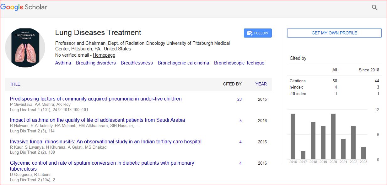
Journal of Lung Diseases & Treatment received 247 citations as per Google Scholar report