Mini Review - (2022) Volume 11, Issue 10
Received: 04-Oct-2022, Manuscript No. ara-22-78289;
Editor assigned: 06-Oct-2022, Pre QC No. P-78289;
Reviewed: 18-Oct-2022, QC No. Q-78289;
Revised: 23-Oct-2022, Manuscript No. R-78289;
Published:
30-Oct-2022
, DOI: 10.37421/2168-9695.2022.11.235
Citation: Zhao, Hongwei. “How the Present Robots Work and Viewpoints for What's To Come.” Adv Robot Autom 11 (2022): 235.
Copyright: © 2022 Zhao H. This is an open-access article distributed under the terms of the Creative Commons Attribution License, which permits unrestricted use, distribution, and reproduction in any medium, provided the original author and source are credited.
The wellsprings of data involved by the robots in clinical practice today are, from one viewpoint, clinical pictures, both pre-and intraoperative, and then again, pictures and estimations of position, of three-layered (3D) shapes, and of power. By and large, starting in 1988, X-beam CT was the main imaging strategy used to design automated stereotactic neurosurgery methodology, which were then acted in the CT scanner room. The predominant exhibition of attractive reverberation imaging (X-ray) for cerebrum imaging, as well as the way that a working room is vastly improved adjusted to a medical procedure than the scanner room, drove us to recommend that stereotactic neurosurgical techniques ought to be directed by a robot that can utilize intraoperative X-ray and radiography.
Robots • Radiography • Neurosurgery
Ultrasound in this manner had all the earmarks of being an extremely valuable wellspring of pictures for directing a medical procedure, including techniques including bones [1]. These wellsprings of pictures have since been generally utilized by careful robots. Different sensors, at first produced for PC vision applications, presently assume a significant part in these robots. Three-layered position sensors or trackers (likewise called 3D localizers), for instance, are a fundamental part of all the careful route frameworks introduced in Segment Activity: grouping of careful robots. Their rule is to make it conceivable to follow the situation in space of specially appointed markers that can be emphatically connected to careful apparatuses or to certain organs [2].
All the while following no less than three such markers unequivocally connected to a similar device makes it conceivable, after an adjustment stage, to decide the place of the instrument's tip as well as its direction. We executed such optical frameworks in clinical practice as soon as 1994; they made it conceivable to follow unbending careful devices and organs — their positions and their developments continuously with an accuracy of short of what one millimeter and short of what one degree. Current frameworks of careful route generally utilize fundamentally the same as optical 3D localizers [3]. Attractive 3D position sensors can be utilized in a few clinical applications; they don't need that the space between the sensor and the item followed be vacant, however they are exceptionally delicate to bothers made by ferromagnetic masses and by a few electric instruments. They can subsequently be challenging to use in the working suite.
The state of the organs that are the object of the intercession is in many cases a fundamental variable. This shape can, obviously, be gotten from preoperative pictures, CT, X-ray, or even ultrasound [4]. Regardless, at times, (directing transpedicular screw obsession, implantation of a complete knee prosthesis, or knee ligamentoplasty, for instance), requesting such an assessment would essentially burden the methodology. A last class of data utilized by careful robots is estimations of mechanical pressure. Estimation of the pressure applied by the administrator on the distal pieces of the robot permits the framework to comprehend the heading where the administrator needs to move the robot, which accordingly goes with the administrator's developments. This guideline has since been utilized in various different robots [5].
One point in like manner in the utilization of this large number of sensors is their alignment. We delineate the significance of this stage with the case of the utilization of computerized picture intensifiers. The pictures they produce are developed as a capability not just of markers like the distance between the item and the locator, yet in addition of the direction of the intensifier comparable to the world's attractive field [6]. These bends are not especially troublesome for undertakings like directing the presentation of a screw towards its objective, the length of the screw and the organs go through similar mutilations. Then again, if we need to take advantage of computerized information from the pictures hence delivered to direct the robot, these contortions should totally be revised. Various adjustment methodologies have in this way been intended to change the spatial directions read on the pictures into solid 3D directions.
A fundamental stage in many applications executing careful robots is the enlistment stage and specifically the matching of each article's direction framework [7]. That is, we have seen that numerous wellsprings of data are obtained, each with its own arrangement of mathematical directions. We will see that the robots additionally have their own such framework. The main framework that intrigues the specialist in the working suite is that of the patient's objective organs and any physical hindrances. It was in this manner important to plan strategies to match these frameworks. These articles can be markers put on the patient. Regardless, with the exception of when the obsession is unbending (stereotactic outline tightens the skull table, or a bone screw for Robodoc), these strategies need accuracy since relative development between the skin and the physical objective remaining parts feasible (for instance, for dots stuck to the patient's skin) [8]. We thusly really like to utilize physical items and, for this situation, to combine the direction frameworks, by looking at, for instance, the region of a vertebra removed from a progression of CT cuts with a disperse plot digitized by palpation of a similar vertebra during the method.
This approach in any case requires the capacity to portion the designs of interest naturally or at least, to handle the pictures to isolate the object of interest from the remainder of the picture. This is generally difficult to do consequently, on the grounds that the difference between the article and the foundation is now and again very low, and on the grounds that the items are some of the time extremely near each other (the articular surfaces of the vertebrae, for instance). This issue of the division of clinical pictures is the subject of a plentiful writing that is difficult to refer to here [9].
The second significant stage is the preparation of the method. It includes characterizing the ideal system, considering the anticipated results of the technique. Two normal cases happen: preoperative preparation, in light of preoperative pictures, and intraoperative preparation, regardless of the utilization of preoperative pictures. Robodoc is one of the best instances of preoperative preparation; its product part, Orthodoc, permits the specialist to pick the prosthesis with a femoral part nearest to the state of the femoral medullary channel, and afterward to choose how to embed it and to characterize the piece of the medullary waterway to machine down to fit the prosthesis stem [10].
For percutaneous cuts, arranging is restricted to permitting the client to pick intuitively the best direction to arrive at the objective while keeping away from perilous regions. Most frequently, a solitary imaging methodology, in cuts, gives all the relevant data. At times, utilizing deduced information is essential. This is the situation, for instance, for stereotactic neurosurgery, where neuroanatomical chart books supply signs for the basal ganglia that the most developed X-ray can't give. A specific issue here is flexible coordinating, which makes it conceivable to twist these chart books to adjust them to the patient's life systems and consequently to design the strategy considering this deduced information.
Google Scholar, Crossref, Indexed at
Google Scholar, Crossref, Indexed at
Google Scholar, Crossref, Indexed at
Google Scholar, Crossref, Indexed at
Google Scholar, Crossref, Indexed at
Google Scholar, Crossref, Indexed at
Google Scholar, Crossref, Indexed at
Google Scholar, Crossref, Indexed at
Google Scholar, Crossref, Indexed at
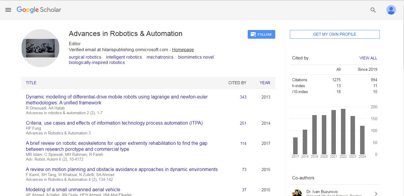
Advances in Robotics & Automation received 1275 citations as per Google Scholar report