Short Communication - (2022) Volume 12, Issue 5
Received: 11-May-2022, Manuscript No. MCCR-22-70298;
Editor assigned: 13-May-2022, Pre QC No. P-70298;
Reviewed: 18-May-2022, QC No. Q-70298;
Revised: 23-May-2022, Manuscript No. R-70298;
Published:
28-May-2022
, DOI: 10.37421/2161-0444.2022.12.624
Citation: Xu, Qidong. “Drug Development and Cancer Epigenetic
Chemical Biology.” Med Chem 12 (2022): 624.
Copyright: © 2022 Xu Q. This is an open-access article distributed under the
terms of the creative commons attribution license which permits unrestricted use,
distribution and reproduction in any medium, provided the original author and
source are credited.
The term "epigenetics" describes how cells and other animals can modify their phenotypes without altering their genomic makeup. Such phenotypic effects are caused by modifications in the transcriptional programme of cells, or the specifics of which genes in a cell are actively transcribed and which are silent, at the molecular level. A group of enzymes and recognition proteins collectively referred to as the chromatinmodifying proteins facilitate genespecific modifications in chromatin structure, which affect the spatiotemporal regulation of the transcriptional programme of a cell. The mixture of DNA and histone proteins known as chromatin is what gives chromosomes their distinctive structural makeup. The three distinct biochemical mechanisms by which the CMPs primarily regulate chromatin conformation in mammals are: methylation of cytosine bases at the C5 position within chromosomal DNA's CpG islands; post-translational modification of the histone proteins that form the core structures of nucleosomes (the spool-like structural units of histone core proteins around which portions of the chromosomal DNA is wound) and exchanges of the histone bases [1].
Due to observations of increased C5-methylation of tumour suppressor genes in some cancers, somatic mutations of DNMT3A in acute myeloid leukaemia (AML), myelodysplastic syndrome (MDS), T cell leukaemia and lymphoma, and loss-of-function mutations in the TET enzymes in AML, chronic lymphocytic leukaemia (CLL), and MDS, the DNA methyltransferase (DNMT) family Decitabine (Dacogen) and azacitidine are the two medications that have been authorised to far for the treatment of MDS that target the DNMT reaction pathway (Vidaza). Guadecitabine (SGI-110), a third inhibitor, is presently undergoing phase 3 studies. These nucleoside-based inhibitors work by being incorporated into the DNA, then covalently trapping the DNMTs and reducing DNA methylation. Protein methyltransferases (PMTs), a broad enzyme class, divide into two families based on the structure of the enzyme active-site: the protein lysine methyltransferases (PKMTs) and the protein arginine methyltransferases, which methylate the lysine and arginine residues on histone proteins (PRMTs). S-adenosylmethionine (SAM) is used by both PKMTs and PRMTs as an all-purpose methyl donor, and both enzymes transfer this methyl group to the side-chain terminal nitrogen of lysine or arginine, respectively. For both families, the reaction process entails the direct transfer of the methyl group from the enzyme-bound SAM to the enzymebound substrate as well as the formation of a ternary enzyme-SAM-substrate complex [2,3].
Additionally to SAM or substrate-competitive inhibitors, substances that bind PRMT3 at a new allosteric location have been described. Additionally, numerous PMTs exhibit sizable dynamic changes in protein structure along their typical catalytic reaction pathway. In order to engage novel recognition elements that develop as a result of protein conformational changes, these structural alterations can occasionally be used in the design of inhibitors. This is perhaps best demonstrated by a group of amino-nucleoside inhibitors of the DOT1L enzyme. Early members of this class were intended to mimic S-adenosylhomocysteine (SAH), a reaction product, and as was predicted, they showed competitive inhibition against SAM. Six separate histone methyltransferases, collectively known as the trithorax group, are in charge of the histone mark H3K4 methylation, which is frequently linked to transcriptional activation in contrast to PRC2-mediated gene repression. MLL1 (KMT2A) and MLL4 (KMT2B), which catalyse H3K4me3 at bivalent promoters, MLL2 (KMT2D) and MLL3 (KMT2C), which catalyse H3K4me1 at enhancers, and SETD1A/SETD1B, which are in charge of a more widespread deposition of H3K4 trimethylation in active chromatin, are members of this family It is not unexpected that these histone marks are commonly altered in cancer and have been linked to both oncogenic and tumour suppressive functions given the significance of these histone marks in gene activation. AML typically exhibits MLL1 fusions with pTEF complex members, which results in inappropriate activation of the carcinogenic RUNX1 gene programme [4,5].
The H3K36 mono- and dimethylation, a histone mark primarily connected to active transcription, is carried out by the NSD family of HMTs, while it has also been demonstrated to be involved in a number of other processes, including alternative splicing and DNA repair. It has been established that every member of this family has an oncogenic role. NSD2, sometimes referred to as WHSC1 or MMSET, is frequently translocated to the immunoglobulin H super-enhancer in multiple myeloma, which causes severe overexpression of NSD2 and correspondingly increased levels of H3K36me2. Hotspot mutations in NSD2 at E1099K have been found in ALL, and they also cause high levels of H3K36me2. AML has been associated with NSD1 or NSD3 fusions with NUP98. The two families that make up the methyl-lysine readers (KMe readers) are the PHD zinc-finger domains and the "Royal Family," which includes the Tudor, Agenet, MBT, chromodomain, and PWWP domains. Recently, it was discovered that a different protein domain, the bromo adjacent homology (BAH) domain of the ORC1 protein, is likewise an H4K20 dimethylation-specific KMe reader. Although these protein families are less likely to have genetic defects than the HMTs, DNMTs, or KDMs, they can occasionally be discovered as translocation partners in a variety of uncommon malignancies. For example, PHF1 fuses with several partners in endometrial stromal sarcoma and ossifying fibromyxoid tumours, while PHF23 fuses with NUP98 in AML [1,2].
In the near future, the findings of several of these trials of investigational CMP-targeted medications will be published, and it will be interesting to observe how these drugs perform in terms of efficacy and safety in humans. It will be crucial to comprehend how these various CMP modulators can be used with one another, with current standard-of-care medicines, with new medications, and with other therapeutic modalities for the efficient control of cancer in addition to their usage as monotherapies. Beyond these achievements, there are still many of chances to use chemical biology in the CMPs. There is currently a shortage of powerful, selective, clinically viable inhibitors for a number of CMP targets that are extremely relevant to particular types of human cancer.
None.
The author reported no potential conflict of interest.
Google Scholar, Crossref, Indexed at
Google Scholar, Crossref, Indexed at
Google Scholar, Crossref, Indexed at
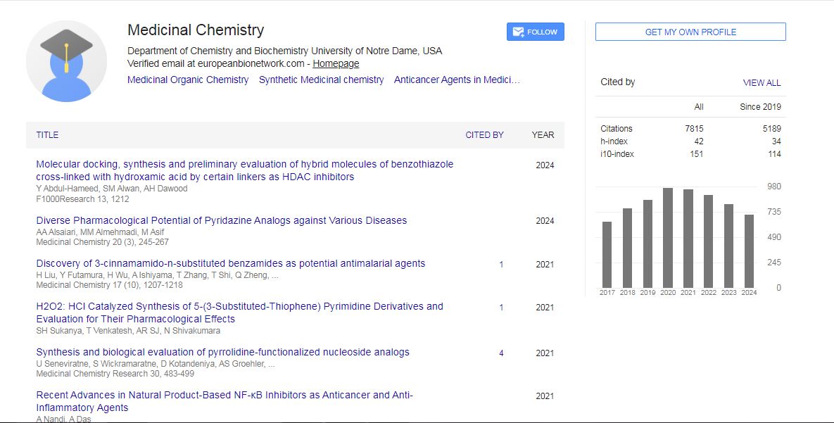
Medicinal Chemistry received 6627 citations as per Google Scholar report