Commentary - (2021) Volume 5, Issue 4
Received: 08-Jul-2021
Published:
22-Jul-2021
, DOI: 10.37421/jmbp.2021.5.124
Citation: Xi-he Zhang. "Components of Histopathology Report." J Microbiol Pathol 5 (2021): 124.
Copyright: �??�?�© 2021 Xi-he Zhang. This is an open-access article distributed under the terms of the Creative Commons Attribution License, which permits unrestricted use, distribution, and reproduction in any medium, provided the original author and source are credited
The National Cancer Institute characterizes histopathology as "the investigation of ailing cells and tissues utilizing a magnifying instrument". Histology is the investigation of tissues, and pathology is the investigation of infection. So taken together, histopathology in a real sense implies the investigation of tissues as identifies with infection. A histopathology report is in some cases called a biopsy report or a pathology report.
The expert specialist who does the assessment under the magnifying lens is known as a pathologist. The tissue that is considered comes from a biopsy or surgery whereby an example of the speculate tissue is chosen and shipped off the research facility. It is then handled and cut into exceptionally slim layers (called areas), stained, and inspected under magnifying instruments to describe the subtleties of the phones in the tissue. For certain infections, the specialist can get an example of the tissue deciphered rapidly using frozen areas. Frozen areas or cuts are utilized sparingly in lymphoma, be that as it may, because of issues in translation and examining. In lymphomas, lymph hubs are the tissue most normally analyzed in histopathology. For some sorts of blood tumors, a bone marrow biopsy may likewise be needed for an authoritative conclusion.
Histopathology provides details regarding careful malignant growth examples are getting increasingly intricate. They may include:
• The tiny appearance of the elaborate tissue
• Special stains
• Molecular strategies
• Other tests
Atomic procedures allude to the capacity to break down cells and tissues at the sub-atomic level, which is at the degree of proteins, receptors, and the qualities that code for these things.
Groundwork for histology
The tissue is then ready for review under a magnifying instrument utilizing either compound obsession or frozen area. In the event that a huge example is given for example from a surgery then a pathologist takes a gander at the tissue test and chooses the part destined to yield a helpful and exact analysis - this part is eliminated for assessment in a cycle ordinarily known as earning or cut up. Bigger examples are sliced to effectively arrange their anatomical constructions in the tape. Certain examples (particularly biopsies) can go through agar pre-installing to guarantee right tissue direction in tape and then, at that point in the square and then, at that point on the demonstrative microscopy slide. This is then positioned into a plastic tape for the greater part of the remainder of the cycle.
1) Chemical obsession
2) Processing
3) Frozen segment preparing
Staining of prepared histology slides
Generally utilized stain in histology is a blend of hematoxylin and eosin (regularly contracted H&E). Hematoxylin is utilized to stain cores blue, while eosin stains cytoplasm and the extracellular connective tissue grid pink. Different mixtures used to shading tissue areas incorporate safranin, Oil Red O, Congo red, silver salts and counterfeit colors.
The histological slides are inspected under a magnifying instrument by a pathologist, a therapeutically qualified expert who has finished a perceived preparing program. This clinical finding is detailed as a pathology report portraying Y.
The histological discoveries and the assessment of the pathologist. On account of malignant growth, this addresses the tissue analysis needed for most treatment conventions. In the expulsion of malignant growth, the pathologist will demonstrate whether the careful edge is cleared, or is included (lingering disease is abandoned). This is finished utilizing either the bread loafing or CCPDMA strategy for handling. Minute visual antiques can conceivably cause misdiagnosis of tests.
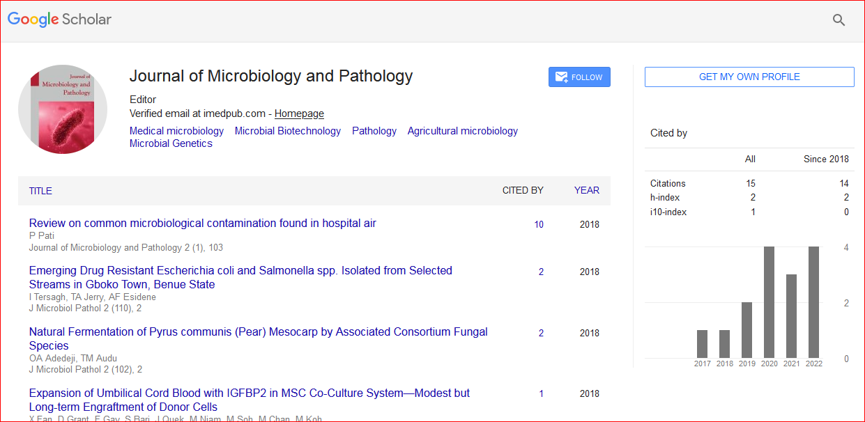
Journal of Microbiology and Pathology received 18 citations as per Google Scholar report