Case Report - (2020) Volume 5, Issue 3
Received: 08-Jun-2020
Published:
28-Jul-2020
, DOI: 10.37421/2573-0312.2020.5.184
Citation: Shimas Salih. “Bony Spurs in a below Knee Amputation: Case Review.” Physiother Rehabil 5 (2020):184. doi: 10.37421/jppr.2020.05.184
Copyright: © 2020 Salih S. This is an open-access article distributed under the terms of the Creative Commons Attribution License, which permits unrestricted use, distribution, and reproduction in any medium, provided the original author and source are credited.
Background
Poor wound healing following a major lower limb amputation can result in poor rehabilitation outcomes, which can be further worsened if the patient has recurrent falls and stump trauma during the wound healing stage. Direct trauma to the stump can result in bony fractures at the margins of the residual bones, and loose bony fragments may form inside the stump. These bony fragments can hinder the process of stump wound healing and interfere with prosthetic limb use and rehabilitation.
Case Description and Methods
We report this unusual case of 54 years old female patient who had a poor postoperative stump wound healing following a direct stump trauma due to a fall during the early postoperative recovery phase. She failed to respond to several weeks of conservative treatment.
Findings
A plain radiograph of the stump showed several body fragments migrated from a fracture at the distal tibial margin. A few weeks later, the patient noticed a sizeable body fragment spontaneously fell from the unhealed area of the stump. Shortly following this, her incisional wound showed signs of complete healing.
Outcomes and Conclusion
We think that this bony spur is the most likely culprit behind the patient's unusually prolonged wound healing. We discussed this case at the local MDT meeting in order to share the knowledge amongst the team and to raise vigilance about the need for early investigations in similar scenarios, particularly following direct trauma to the amputation residuum.
Amputation • Trauma • Rehabilitation
Latest data published by Public Health England 2019 showed that the number of transtibial amputations has been on the rise over the last few years with 7545 new amputations between 2015-2018 compared with 6957 between 2012-2015 [1]. Several medical conditions predispose to major lower limb amputations, for example, atherosclerotic peripheral vascular disease (PVD) and poorly controlled Diabetes Mellitus (DM).2 The combination of Diabetes Mellitus and PVD increases the chance of lower limb amputation by 1.5 folds compared to PVD alone [2.3]. In addition to PVD and DM, other risk factors contribute to the incidence of major lower limb amputation are age, obesity, hypertension, dyslipidaemia and smoking [4].
Falls and near misses are some of the frequently observed phenomena in people with lower limb amputation, particularly in the presence of other predisposing factors such as reduced premorbid mobility, frailty, old age, advanced osteoarthritis, soft tissue disease, and poly-pharmacy [5]. Patients with major lower limb amputations have an estimated annual incidence of falls of around 52.4% [6]. A large proportion of those who have not yet sustained a fall reported a significant fear of falling due to lack of confidence [6]. What is even more concerning is that 50% of people with lower-limb amputation report at least one fall in a single year, approximately 40.4% of them incur a degree of injury, and 20% require medical attention [7,8]. Transtibial amputees are at higher risk of noteworthy falls and injuries compared with their transfemoral counterparts. This is because the former group achieve higher-level functions speedier and often indulge themselves in a broader range of activities at an early stage of their rehabilitation, subsequently increase their chance of falling [9,10]. Some studies showed that transtibial amputees who repeatedly fall, tend to achieve a functional walking earlier than those who have not experienced any falls which reflects their commitment to attempt walking [11].
A 54-year female patient underwent left-sided transtibial amputation due to persistent ulcers on her left foot, and toes resulted in necrotising fasciitis. She is a heavy cigarette smoker with a background of insulin dependent Diabetes Mellitus and dyslipidaemia. Ten days following her amputation had a significant mechanical fall at home while was making her way to the bathroom using two crutches. She landed directly on her residuum and incurred a large bruise involving the whole anterior aspect of the residuum. A few days later, she noticed a small area of wound dehiscence at the centre of the surgical scar with a small amount of serous discharge. She had wound care twice a week while simultaneously progressing well with Pneumatic Post-Amputation Mobility Aid training. She completed a seven days course of Flucloxacillin 500mg oral capsules according to the swab culture and sensitivity results which confirmed the growth staphylococcus Aureus bacteria.
Four months later, her wound healed up apart from a central rounded area of approximately 0.5cm x 0.5cm in size. We initially suspected a herniation of the stump myoplasty at the dehiscent point. This unhealed area continued to leak a small amount of haemo-serous fluid, yet there were no features of infection. A few weeks later the patient reported a foreign body sensation inside the wound which she described as ''a bone splinter'' at the distal end of her residuum. A plain radiograph of the stump showed free-floating bony fragments noted at the periphery of the anterior tibial fracture with no features of osteomyelitis.
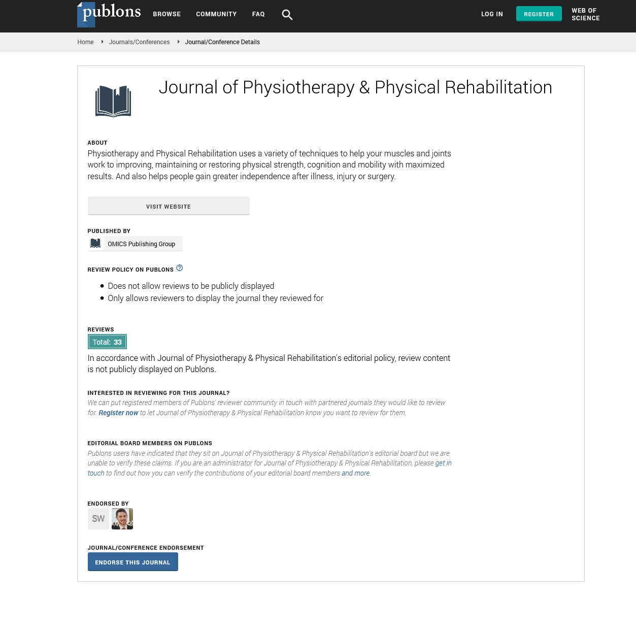
The patient continued to progress with her rehabilitation as planned. She started to use a prosthetic limb for around 3-4 hours a day despite the area at the centre of the scan still not fully healed. Five weeks later, she reported a small piece of bone spontaneously dropped from the open area as shown in Figure 1. (Figure 1)

Figure 1. Two images showing the spontaneously expelled bony fragments.
A week later, she had a medical assessment which confirms complete healing of the operation wound with no residual discharge or dehiscence. A repeat X-ray of the residuum showed that the bony fragments noted on the previous images had disappeared; however, there is still a bony fragment abutting the tibia as shown in Figure 2. (Figure 2)

Figure 2. On the right: A pre-expulsion X-ray showing free-floating bony fragments noted at the periphery of the anterior tibia fracture. on the left: A repeat X- Ray 6 weeks later demonstrated that the bony fragments noted on the previous imaging distal to the tibial amputation site have been expelled.
One of the commonly observed complications following major lower limb amputation is poor wound healing which is more significant in patients with existing comorbidities such as diabetes and PVD. 12 Also, the incidence of wound infection is higher if the wound healing process is prolonged. The chance of wound infections following a major lower limb amputation is estimated to be between 13% and 40%, determined by the presence of other risk factors such as poorly controlled Diabetes Mellitus, PVD; chronic skin conditions etc [12].
Unhealed wounds associated with other amputation related complications such as phantom limb pain or underlying osteomyelitis often require surgical attention [13].
A Stump revision surgery is a common surgical measure entails converting a transtibial to a transfemoral level in these scenarios [13]. Moreover, delayed healing of the stump wound can be a sign of an underlying pathology hindering the healing process, for example, bony fractures following direct trauma to the stump or osteomyelitis.14 Direct trauma to the stump is often a consequence of falling and landing on the stump. It, sometimes, results in fractures at the margins of the residual bones, floating bony spurs or ectopic bone tissue formation inside the residuum. The patient usually reports a considerable pain which may interfere with the use of the prosthetic limb delaying the progress of the post-amputation rehabilitation [14]. The mechanism of ectopic bone tissue formation around the residual bones is unclear. It is, however, thought to be due to extensive periosteal stripping and retention at the amputation site following the amputation or subsequent trauma to the stump [15]. Large bony spurs approaching the skin of the stump may result in skin pressure, stress and ulceration, they may also lead to persistent sinuses, skin infections and poor wound healing [16].
Persistent infection of the amputation wound should warrant further investigation. High-resolution ultrasound scan helps in identifying pockets of fluid collection inside the stump and underlying inflammatory changes using the Doppler Effect [17]. Plain radiography helps to raise suspicions around underlying osteomyelitis; however, it is the imaging modality of choice to diagnose heterotopic bone tissue, fractures and bone fragments [17]. Magnetic resonance imaging is the investigation of choice which provides details about local effusion, bone spurs, inflammation, osteomyelitis and other soft tissue pathologies. It is also useful to monitor response to conservative management as well as to guide the decision about further treatment options including surgical interventions [17, 18]
The amputation residuum is unable to tolerate high mechanical stress; as a result, it attempts to adapt by process of remodeling, which leads to the development of inflammatory changes involving the stump bones and soft tissues.18 Sometimes this inflammatory response weakens the residual bones leading to stress fractures and fragmentations [18].
We found this case very interesting because it highlights the significance of the relationship between post-amputation traumatic fall and poor wound healing. Tibial end fracture in transtibial amputation is not a commonly reported phenomenon in the literature.
While there are an extensive number of studies about stress fractures of the residual bones, there are not many available regarding fractures following direct trauma to the stump. It is either because it is not a frequently occurring problem, or because post-trauma fractures remain clinically insignificant.
The spontaneously ejected bony fragments in our case are likely to have migrated from the tibial fracture site following the fall. We discussed this case at the local MDT meeting in order to share the knowledge amongst the team about the need for early investigations for similar scenarios in the future, particularly in the presence of direct trauma to the amputation residuum.