Editorial - (2022) Volume 13, Issue 4
Received: 05-Apr-2022, Manuscript No. jhmi-22-62401;
Editor assigned: 07-Apr-2022, Pre QC No. P-62401;
Reviewed: 10-Apr-2022, QC No. Q-62401;
Revised: 15-Apr-2022, Manuscript No. R-62401;
Published:
20-Apr-2022
, DOI: 10.37421/jhmi.2022.13.413
Citation: Khatoon, Reshma. “Attack of SARS-CoV-2 Virus in the Brain.” J Health Med Informat 13 (2022): 413.
Copyright: © 2022 Khatoon R. This is an open-access article distributed under the terms of the Creative Commons Attribution License, which permits unrestricted use, distribution, and reproduction in any medium, provided the original author and source are credited.
Coronavirus disease 2019 (COVID-19) is a heterogeneous and complex disease caused by an abnormal immune response to a human coronavirus infection known as SARS-CoV-2 (severe acute respiratory syndrome coronavirus 2). COVID-19 exhibits a wide spectrum of severity, from asymptomatic to deadly clinical results, according to substantial clinical findings in the fields of diagnosis, therapeutic management, and prevention. SARSCoV- 2 primarily impacts lung function during the acute phase of infection by invading and replicating in the upper respiratory tract, then rapidly migrating to the lower respiratory tracts, resulting in severe or deadly pneumonia. However, a variety of symptoms last a long period after infection and correspond to SARS-effects CoV-2's on multiorgan systems [1].
As indicated by the National Institute for Health and Care Excellence (NICE) rules and reports of the British Office for National Statistics (ONS) the post- COVID-19 circumstances inside the aspiratory, cardiovascular and sensory system, regularly called long-COVID, have been seen in over 20% of people for quite some time following intense SARS-CoV-2 disease. The high biodiversity and irresistible not set in stone by the arrangements of transformations that outcome in variations of explicit contagiousness and antigenicity. In view of SARS-CoV-2 sub-atomic design and hereditary heterogeneity, five "variations of concern" (VoC) have been described by their irresistible potential, spread and casualty rate [2]. These attributes rely upon the changes in viral underlying proteins, including envelope (E), layer (M), nucleocapsid (N), and the spike protein (S) that coordinate replication and viral gathering. In the part of viral science, the variety of these proteins results from the high pace of hereditary varieties and determinates viral pathogenicity, infectivity, and antigenicity. Since the COVID-19 pandemic's beginning, researchers have been attempting to characterize the total component that permits the viral particles to enter and taint solid human cells. SARS-CoV-2, as other β-COVIDS (β-CoVs), takes advantage of the S protein to start the collaboration between viral capsid and the host cell films. The systems basic the section into the host cells enlist the useful areas of S protein that perceive explicit host receptors and intercede the combination with have cell films [3].
This cycle begins from the initiation of film combination movement through the cleavage of S protein into S1 and S2 spaces interceded by the host cell proteases, including Transmembrane Protease Serine 2 (TMPRSS2), furin, and cathepsins. Proteolytic handling inside the S1 subunit is pivotal for acknowledgment and cooperation between its Receptor-restricting Space (RBD) and the host receptors. Both clinical and exploratory examinations have recognized the angiotensin-changing over protein 2 (ACE2) as the section receptor for SARS-CoV-2 into the human cells. As has been shown, the declaration of transmembrane, however not dissolvable ACE2 isoform was decidedly connected with viral RNA load and TMPRSS2 articulation relying upon the age and natural sex of the host. Also, the gamble of contamination of SARS-CoV-2 and advancement of its replication can be advanced by Renin Angiotensin Framework (RAS) modulators, including Angiotensin Receptors Blockers (ARB), as has been exhibited in refined Vero E6 cells [4].
The variety in sickness seriousness after SARS-CoV-2 contamination is directed by the solid cell tropism and the expansive articulation of ACE2 in a few organs of the body, and is subsequently firmly connected to moderate brokenness inside the insusceptible, aspiratory, cardiovascular and sensory systems. Be that as it may, there is expanding proof on the enlistment of the profoundly sialylated cell glycoconjugates in viral section, motor and resistant guideline in SARS-CoV-2 contaminations. Since the underlying examination of the N-terminal area (NTD) of SARS-CoV-2 has revealed the capacity of explicit amino corrosive arrangements to frame sialoside restricting pockets, the connection with the host surface glycocalyx can be practically engaged with the quick infectivity and spread in sialic corrosive rich organs. The tantamount investigation of human β-CoVs in the field of instruments of contamination, tropism, and pathogenesis uncovered the job of sialic acids as controllers of SARS-CoV-2's infective potential and obtrusiveness [5]. This audit momentarily centers around the significance of sialic acids in the SARS-CoV-2-related pathology in the focal sensory system. We present the sialoglycans as an option viral section instrument and examine how human cell surface sialome has been impacted by COVID-19 and relates to clinical aggravations inside the cerebrum.
None.
Google Scholar, Crossref, Indexed at
Google Scholar, Crossref, Indexed at
Google Scholar, Crossref, Indexed at
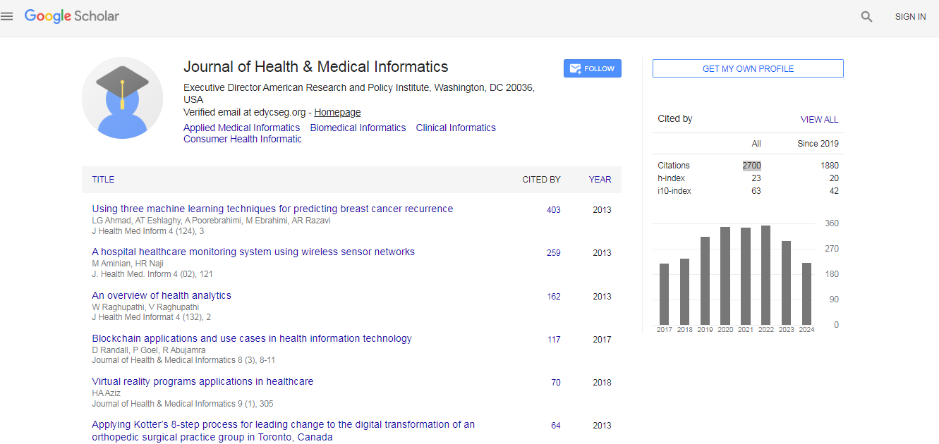
Journal of Health & Medical Informatics received 2700 citations as per Google Scholar report