Editorial - (2022) Volume 12, Issue 3
Received: 04-May-2022, Manuscript No. jpdbd-22-70235;
Editor assigned: 07-May-2022, Pre QC No. P-70235;
Reviewed: 09-May-2022, QC No. Q-70235;
Revised: 14-May-2022, Manuscript No. R-70235;
Published:
19-May-2022
, DOI: 10.37421/2153-0769.2022.12.320
Citation: Siberil, Lucas. “Apoptosis-specific Genes Highlights
Metabolic Status of Hostile Cells in Adenocarcinoma.” Metabolomics 12 (2022):
320.
Copyright: © 2022 Siberil L. This is an open-access article distributed under the
terms of the Creative Commons Attribution License, which permits unrestricted
use, distribution, and reproduction in any medium, provided the original author
and source are credited.
The job of autophagy in cellular breakdowns in the lungs is as yet disputable, predominantly in light of the fact that the perception of autophagy levels in patients stays testing. One intriguing methodology comprises of examining autophagy at the transcriptomic level. Autophagy is a selfdegradative system engaged with numerous natural cycles, including cell demise, endurance, multiplication or relocation. In growths, autophagy assumes a significant part in tumorigenesis as well as disease movement and protection from treatments. Typically, an elevated degree of autophagy in dangerous cells has been related with growth movement and unfortunate prognostic for patients. Nonetheless, the examination of autophagy levels in patients stays troublesome, particularly on the grounds that evaluation of autophagy proteins is trying in the growth microenvironment. Lung growths are among the most well-known on the planet and are one of the main sources of disease passings overall. Among them, NSCLC represents 85% of all lung cancer cases and it is mostly made out of lung squamous cell carcinoma (LUSC) and adenocarcinoma (LUAD). Albeit the accessible helpful weapons store, including surgeries, chemotherapy, designated particles and immunotherapy, have permitted obvious advancement in cellular breakdown in the lungs treatment, the 5-year endurance pace of NSCLC patients stays unsuitable [1- 3]. This overall shortcoming is predominantly because of the absence of data about the cancer microenvironment during therapy choice. Hence, numerous examinations have zeroed in on the revelation of another prognostic appraisal strategy to help individualized treatment of NSLCL patients [4].
Autophagy is a cell cycle related with the pervasiveness and movement of cellular breakdown in the lungs. Autophagy is a rationed catabolism pathway that assumes a critical part in the support of cell homeostasis. It is a multistep component, comprising of the development of the phagophore that extends and immerses designated proteins or organelles in a twofold layer vesicle called the autophagosome, lastly melds with late endosomes as well as lysosomes. This cycle is coordinated by an enormous assortment of proteins, including the autophagic proteins (Atg), coordinated in edifices. Autophagy enlistment is tweaked by two protein edifices, the ULK1/2 (unc51-like autophagy enacting kinase) and the Beclin-1/PI3KC3 (class III phosphatidylinositol 3-kinase) buildings. When actuated, these edifices enlist different proteins engaged with the stretching and development of autophagosomes, including the two formed frameworks Atg12-Atg5-Atg16L and LC3. After fulfillment, the experienced autophagosome wires with lysosomes to shape autolysosomes, wherein the sequestered materials and organelles are debased by lysosomal chemicals. Numerous transcriptomic examinations performed on autophagy qualities have zeroed in on the disclosure of new biomarkers to foresee the productivity of hostile to growth treatments and to direct individualized treatment in NSCLC patients. Notwithstanding, most of these examinations depend on worldwide transcriptomic examination of the entire growth microenvironment, and not many examinations have been done on threatening cells themselves [5].
None.
Google Scholar, Crossref, Indexed at
Google Scholar, Crossref, Indexed at
Google Scholar, Crossref, Indexed at
Google Scholar, Crossref, Indexed at
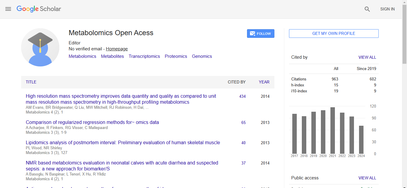
Metabolomics:Open Access received 895 citations as per Google Scholar report