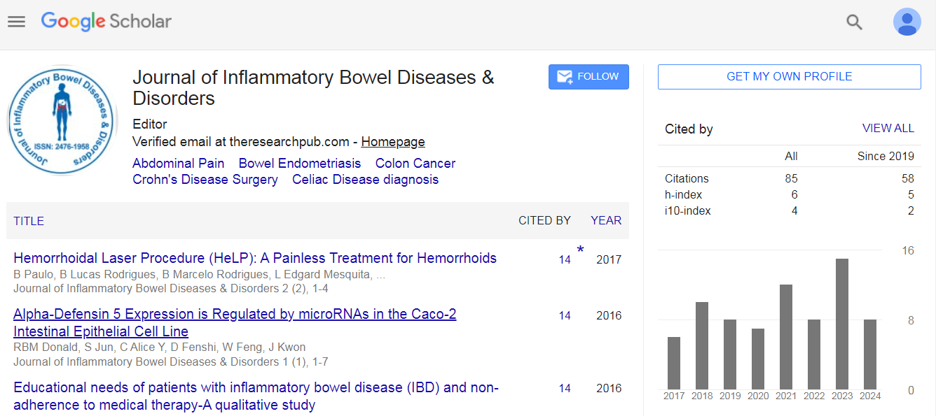
Paulo Boarini, Lucas Rodrigues Boarini, Marcelo Rodrigues Boarini, Edgard Mesquita de Lima and Paulo Azeredo Candelaria
Introduction: Hemorrhoidal disease is associated with the theory of arterial blood hyperflow causing swellings in hemorrhoids and, consequently, hyperplasia and venous congestion. The technique helps to promote the obliteration of the terminals of the superior rectal artery branches without the need for anesthesia by electrofulguration with diode fiber LASER. The objective of this study is to describe the results of 55 patients with hemorrhoidal disease treated by the Hemorrhoidal LASER Procedure technique.
Method: Without the need of anesthesia, terminal arterioles of the upper rectal artery are identified by a Doppler transducer (20 MHz probe 3 mm) placed on a specially designed proctoscope. After identification, it promotes arteriolar electrofulguration at 980 nm fiber laser diode, causing interruption of hemorrhoidal flow. This procedure is repeated circumferentially, following the clockwise positions.
Results: Between 2011 and 2014, 55 patients underwent the Hemorrhoidal LASER Procedure technique for hemorrhoidal disease grades I, II and III. There was no need for anesthesia and only two patients required sedation for the procedure. The overall satisfaction rate was 89%, with symptom resolution in 84% and a decrease of at least one grade in hemorrhoidal disease in 80% of cases.
Conclusion: Hemorrhoidal LASER Procedure is a painless outpatient technique that does not require anesthesia, in addition to being safe and easy to perform. It is effective in reducing symptoms and complications of the hemorrhoidal disease grades I and II, with high satisfaction rates.
A. Aomari, M. Firwana, A. Rahaoui, Kh. Abdelwali, I. Benelbarhdadi and FZ Ajana
Background: The aminosalicylates have a direct local anti-inflammatory effect on the mucous membrane of the small intestine and colon. They have been used for many years in the treatment of inflammatory bowel disease (IBD), these are, generally, well tolerated, however, like all drugs; they may, in rare cases, cause Side effects. We report a patient with a distal ulcerative colitis who presented with acute pancreatitis under Pentasa® enema.
Conclusion: The evolution was marked by the disappearance of pain and vomiting with normalization of lipase and enteral nutrition was retained without any difficulties. The diagnosis is pancreatitis in 5-ASA. The acute pancreatitis secondary to aminosalicylates is a very rare complication. In our case, taking the Pentasa® enema for 03 days was sufficient to cause acute inflammation of the pancreas.
Cordesmeyer A, Buhr J, Hoffmann MW and Allemeyer EH
Kazuhito Sasaki, Mikiya Takao, Fuyo Yoshimi, Keisuke Hata, Toshiaki Watanabe and Tatsuo Iijima
Ulcerative colitis (UC) is a form of inflammatory bowel disease that typically involves the colorectum; it has been reported that a certain proportion of patients with UC also develop ileitis, leading to ileal perforation in very extreme cases. We report a 66-year-old male with UC who presented with ileal perforation eight days after proctocolectomy. Although this situation is very rare, differential diagnoses for small bowel perforation after UC surgery could include backwash ileitis, cytomegalovirus (CMV) infection, Crohn’s disease, diffuse enteritis, ischemic enteritis, Behçet’s disease, medication adverse effect, and iatrogenic injury. Of these, backwash ileitis or diffuse enteritis is the most probable diagnosis in our case. Granulomas and transmural lymphoid aggregates with associated mucosal ulceration were absent. In addition, no signs or symptoms suggestive of Crohn’s disease were seen postoperatively. Thus, the original diagnosis was likely fulminant UC. Infectious enteritis (including CMV), ischemic enteritis, and Behçet’s diseases were clinically ruled out. Stool cultures and CMV antigen testing were negative. Moreover, histopathology revealed no evidence of CMV infection. Only a few cases of ileal perforation after UC surgery have been reported thus far. Surgeons should evaluate for perforation of the small bowel intraoperatively. Resection of the affected ileum is still a matter of debate. Although the inflammation is usually reversible and preservation of the distal ileum is vital for the creation of an ileal pouch and the avoidance of high output, the rare possibility of ileal perforation should be kept in mind in extreme cases of fulminant UC.
Martin Bosch, Franc H. Hetzer and Diana Effinger-Sehmer

 Spanish
  Chinese
  Russian
  German
  French
  Japanese
  Portuguese
  Hindi
Hindi 